 Movie
Movie Controller


+ Open data

- Basic information
| Entry | Database: PDB / ID: 8ayc | ||||||
|---|---|---|---|---|---|---|---|
| Title | Scalindua brodae amxFabZ, I69G mutant | ||||||
 Components Components | Beta-hydroxyacyl-(Acyl-carrier-protein) dehydratase | ||||||
 Keywords Keywords | LIPID BINDING PROTEIN / fatty acid biosynthesis | ||||||
| Function / homology |  Function and homology information Function and homology information3-hydroxyacyl-[acyl-carrier-protein] dehydratase / (3R)-hydroxyacyl-[acyl-carrier-protein] dehydratase activity / lipid A biosynthetic process / membrane / cytoplasm Similarity search - Function | ||||||
| Biological species |  Candidatus Scalindua brodae (bacteria) Candidatus Scalindua brodae (bacteria) | ||||||
| Method |  X-RAY DIFFRACTION / SYNCHROTRON / FOURIER SYNTHESIS / Resolution: 1.75 Å X-RAY DIFFRACTION / SYNCHROTRON / FOURIER SYNTHESIS / Resolution: 1.75 Å | ||||||
 Authors Authors | Dietl, A. / Barends, T. | ||||||
| Funding support | European Union, 1items
| ||||||
 Citation Citation | Journal: J.Biol.Chem. / Year: 2023 Title: Structures of an unusual 3-hydroxyacyl dehydratase (FabZ) from a ladderane-producing organism with an unexpected substrate preference. Authors: Dietl, A. / Wellach, K. / Mahadevan, P. / Mertes, N. / Winter, S.L. / Kutsch, T. / Walz, C. / Schlichting, I. / Fabritz, S. / Barends, T.R.M. | ||||||
| History |
|
- Structure visualization
| Structure viewer | Molecule: MolmilJmol/JSmol |
|---|
- Downloads & links
-Download
| PDBx/mmCIF format | 8ayc.cif.gz | 78.1 KB | Display | PDBx/mmCIF format |
|---|---|---|---|---|
| PDB format | pdb8ayc.ent.gz | 48.7 KB | Display | PDB format |
| PDBx/mmJSON format | 8ayc.json.gz | Tree view | PDBx/mmJSON format | |
| Others |  Other downloads Other downloads |
-Validation report
| Arichive directory | https://data.pdbj.org/pub/pdb/validation_reports/ay/8aycftp://data.pdbj.org/pub/pdb/validation_reports/ay/8ayc | HTTPS FTP |
|---|
-Related structure data
| Related structure data |  7qv0C 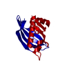 8aybC  8aydC  8ayiC  1u1zS S: Starting model for refinement C: citing same article ( |
|---|---|
| Similar structure data |
-Links
 PDBj
PDBj
- Assembly
| Deposited unit | 
| ||||||||||||
|---|---|---|---|---|---|---|---|---|---|---|---|---|---|
| 1 | x 6
| ||||||||||||
| Unit cell |
| ||||||||||||
| Components on special symmetry positions |
|
-Components
| #1: Protein | Mass: 15498.063 Da / Num. of mol.: 1 Source method: isolated from a genetically manipulated source Details: Scalindua brodae amxFabZ I69G mutant / Source: (gene. exp.) Candidatus Scalindua brodae (bacteria) / Gene: SCABRO_02230 / Production host: |
|---|---|
| #2: Water | ChemComp-HOH /  Mass: 18.015 Da / Num. of mol.: 54 / Source method: isolated from a natural source / Formula: H2O Mass: 18.015 Da / Num. of mol.: 54 / Source method: isolated from a natural source / Formula: H2O |
-Experimental details
-Experiment
| Experiment | Method: X-RAY DIFFRACTION / Number of used crystals: 1 |
|---|
- Sample preparation
| Crystal | Density Matthews: 2.71 Å3/Da / Density % sol: 54.62 % |
|---|---|
| Crystal grow | Temperature: 293 K / Method: vapor diffusion, hanging drop / Details: 1.0 M Sodium formate |
-Data collection
| Diffraction | Mean temperature: 100 K / Serial crystal experiment: N |
|---|---|
| Diffraction source | Source: SYNCHROTRON / Site: SLS  / Beamline: X10SA / Wavelength: 1.00001 Å / Beamline: X10SA / Wavelength: 1.00001 Å |
| Detector | Type: DECTRIS PILATUS 6M / Detector: PIXEL / Date: Aug 22, 2022 |
| Radiation | Protocol: SINGLE WAVELENGTH / Monochromatic (M) / Laue (L): M / Scattering type: x-ray |
| Radiation wavelength | Wavelength: 1.00001 Å / Relative weight: 1 |
| Reflection | Resolution: 1.746→39.39 Å / Num. obs: 15812 / % possible obs: 92.2 % / Redundancy: 5.7 % / Biso Wilson estimate: 46.21 Å2 / CC1/2: 0.999 / Rmerge(I) obs: 0.042 / Net I/σ(I): 19.1 |
| Reflection shell | Resolution: 1.746→1.817 Å / Rmerge(I) obs: 0.876 / Num. unique obs: 793 / CC1/2: 0.423 |
- Processing
| Software |
| ||||||||||||||||||||||||||||||||||||||||||||||||||||||||||||||||||||||||||||||||||||
|---|---|---|---|---|---|---|---|---|---|---|---|---|---|---|---|---|---|---|---|---|---|---|---|---|---|---|---|---|---|---|---|---|---|---|---|---|---|---|---|---|---|---|---|---|---|---|---|---|---|---|---|---|---|---|---|---|---|---|---|---|---|---|---|---|---|---|---|---|---|---|---|---|---|---|---|---|---|---|---|---|---|---|---|---|---|
| Refinement | Method to determine structure: FOURIER SYNTHESIS Starting model: 1U1Z Resolution: 1.75→39.39 Å / SU ML: 0.2071 / Cross valid method: FREE R-VALUE / σ(F): 1.35 / Phase error: 23.7774 Stereochemistry target values: GeoStd + Monomer Library + CDL v1.2
| ||||||||||||||||||||||||||||||||||||||||||||||||||||||||||||||||||||||||||||||||||||
| Solvent computation | Shrinkage radii: 0.9 Å / VDW probe radii: 1.11 Å / Solvent model: FLAT BULK SOLVENT MODEL | ||||||||||||||||||||||||||||||||||||||||||||||||||||||||||||||||||||||||||||||||||||
| Displacement parameters | Biso mean: 54.89 Å2 | ||||||||||||||||||||||||||||||||||||||||||||||||||||||||||||||||||||||||||||||||||||
| Refinement step | Cycle: LAST / Resolution: 1.75→39.39 Å
| ||||||||||||||||||||||||||||||||||||||||||||||||||||||||||||||||||||||||||||||||||||
| Refine LS restraints |
| ||||||||||||||||||||||||||||||||||||||||||||||||||||||||||||||||||||||||||||||||||||
| LS refinement shell |
| ||||||||||||||||||||||||||||||||||||||||||||||||||||||||||||||||||||||||||||||||||||
| Refinement TLS params. | Method: refined / Origin x: -1.95733847078 Å / Origin y: 21.8162410012 Å / Origin z: 5.52083718642 Å
| ||||||||||||||||||||||||||||||||||||||||||||||||||||||||||||||||||||||||||||||||||||
| Refinement TLS group | Selection details: (chain 'A' and resid 1 through 141) |
