 Movie
Movie Controller


+ Open data

- Basic information
| Entry | Database: PDB / ID: 8ay4 | ||||||
|---|---|---|---|---|---|---|---|
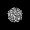
| Title | Human rhinovirus 2 virion in situ | ||||||
 Components Components |
| ||||||
 Keywords Keywords | VIRUS / Human rhinovirus 2 / in situ / cryo-EM. | ||||||
| Function / homology |  Function and homology information Function and homology informationsymbiont-mediated suppression of host cytoplasmic pattern recognition receptor signaling pathway via inhibition of RIG-I activity / picornain 2A / symbiont-mediated suppression of host mRNA export from nucleus / symbiont genome entry into host cell via pore formation in plasma membrane / picornain 3C / T=pseudo3 icosahedral viral capsid / host cell cytoplasmic vesicle membrane / ribonucleoside triphosphate phosphatase activity / nucleoside-triphosphate phosphatase / channel activity ...symbiont-mediated suppression of host cytoplasmic pattern recognition receptor signaling pathway via inhibition of RIG-I activity / picornain 2A / symbiont-mediated suppression of host mRNA export from nucleus / symbiont genome entry into host cell via pore formation in plasma membrane / picornain 3C / T=pseudo3 icosahedral viral capsid / host cell cytoplasmic vesicle membrane / ribonucleoside triphosphate phosphatase activity / nucleoside-triphosphate phosphatase / channel activity / monoatomic ion transmembrane transport / DNA replication / RNA helicase activity / endocytosis involved in viral entry into host cell / symbiont-mediated activation of host autophagy / RNA-directed RNA polymerase / cysteine-type endopeptidase activity / viral RNA genome replication / RNA-directed RNA polymerase activity / virion attachment to host cell / host cell nucleus / DNA-templated transcription / structural molecule activity / proteolysis / RNA binding / zinc ion binding / ATP binding Similarity search - Function | ||||||
| Biological species |  rhinovirus A2 rhinovirus A2 | ||||||
| Method | ELECTRON MICROSCOPY / single particle reconstruction / cryo EM / Resolution: 4.7 Å | ||||||
 Authors Authors | Ishemgulova, A. / Mukhamedova, L. / Trebichalska, Z. / Payne, P. / Smerdova, L. / Moravcova, J. / Hrebik, D. / Buchta, D. / Skubnik, K. / Fuzik, T. ...Ishemgulova, A. / Mukhamedova, L. / Trebichalska, Z. / Payne, P. / Smerdova, L. / Moravcova, J. / Hrebik, D. / Buchta, D. / Skubnik, K. / Fuzik, T. / Novacek, J. / Plevka, P. | ||||||
| Funding support |  Czech Republic, 1items Czech Republic, 1items
| ||||||
 Citation Citation | Journal: To Be Published Title: Endosome rupture enables enteroviruses to infect cells. Authors: Ishemgulova, A. | ||||||
| History |
|
- Structure visualization
| Structure viewer | Molecule: MolmilJmol/JSmol |
|---|
- Downloads & links
-Download
| PDBx/mmCIF format | 8ay4.cif.gz | 263.1 KB | Display | PDBx/mmCIF format |
|---|---|---|---|---|
| PDB format | pdb8ay4.ent.gz | 215.7 KB | Display | PDB format |
| PDBx/mmJSON format | 8ay4.json.gz | Tree view | PDBx/mmJSON format | |
| Others |  Other downloads Other downloads |
-Validation report
| Arichive directory | https://data.pdbj.org/pub/pdb/validation_reports/ay/8ay4ftp://data.pdbj.org/pub/pdb/validation_reports/ay/8ay4 | HTTPS FTP |
|---|
-Related structure data
| Related structure data | 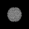 15710MC 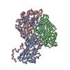 8ay5C M: map data used to model this data C: citing same article ( |
|---|---|
| Similar structure data |
-Links
 PDBj
PDBj

- Assembly
| Deposited unit | 
|
|---|---|
| 1 | x 60
|
-Components
| #1: Protein | Mass: 30645.367 Da / Num. of mol.: 1 / Source method: isolated from a natural source / Source: (natural) rhinovirus A2Plasmid details: Rhinovirus 2 is initially purified and used for infection of cells. Micrographs were collected on lamellipodia of infected Cos7 cells. References: UniProt: P04936 |
|---|---|
| #2: Protein | Mass: 27899.426 Da / Num. of mol.: 1 / Source method: isolated from a natural source / Source: (natural) rhinovirus A2Plasmid details: Rhinovirus 2 is initially purified and used for infection of cells. Micrographs were collected on lamellipodia of infected Cos7 cells. References: UniProt: P04936 |
| #3: Protein | Mass: 26107.793 Da / Num. of mol.: 1 / Source method: isolated from a natural source / Source: (natural) rhinovirus A2Plasmid details: Rhinovirus 2 is initially purified and used for infection of cells. Micrographs were collected on lamellipodia of infected Cos7 cells. References: UniProt: P04936 |
| #4: Protein/peptide | Mass: 2810.063 Da / Num. of mol.: 1 / Source method: isolated from a natural source / Source: (natural) rhinovirus A2Plasmid details: Rhinovirus 2 is initially purified and used for infection of cells. Micrographs were collected on lamellipodia of infected Cos7 cells. |
-Experimental details
-Experiment
| Experiment | Method: ELECTRON MICROSCOPY |
|---|---|
| EM experiment | Aggregation state: PARTICLE / 3D reconstruction method: single particle reconstruction |
- Sample preparation
| Component | Name: rhinovirus A2 / Type: VIRUS / Entity ID: all / Source: NATURAL |
|---|---|
| Source (natural) | Organism: rhinovirus A2 |
| Details of virus | Empty: NO / Enveloped: NO / Isolate: OTHER / Type: VIRION |
| Buffer solution | pH: 7.4 |
| Specimen | Embedding applied: NO / Shadowing applied: NO / Staining applied: NO / Vitrification applied: YES |
| Specimen support | Grid material: GOLD / Grid mesh size: 200 divisions/in. / Grid type: Quantifoil R2/1 |
| Vitrification | Instrument: FEI VITROBOT MARK IV / Cryogen name: ETHANE / Humidity: 100 % |
- Electron microscopy imaging
| Experimental equipment |  Model: Titan Krios / Image courtesy: FEI Company |
|---|---|
| Microscopy | Model: FEI TITAN KRIOS |
| Electron gun | Electron source:  FIELD EMISSION GUN / Accelerating voltage: 300 kV / Illumination mode: OTHER FIELD EMISSION GUN / Accelerating voltage: 300 kV / Illumination mode: OTHER |
| Electron lens | Mode: BRIGHT FIELD / Nominal magnification: 105000 X / Nominal defocus max: 3000 nm / Nominal defocus min: 1800 nm / Cs: 2.7 mm |
| Specimen holder | Cryogen: NITROGEN / Specimen holder model: FEI TITAN KRIOS AUTOGRID HOLDER |
| Image recording | Average exposure time: 10 sec. / Electron dose: 50 e/Å2 / Film or detector model: GATAN K2 QUANTUM (4k x 4k) |
| EM imaging optics | Energyfilter name: GIF Bioquantum / Energyfilter slit width: 20 eV |
| Image scans | Movie frames/image: 40 / Used frames/image: 1-40 |
- Processing
| Software | Name: UCSF ChimeraX / Version: 1.4/v9 / Classification: model building / URL: https://www.rbvi.ucsf.edu/chimerax/ / Os: macOS / Type: package | ||||||||||||||||||||||||||||||||||||||||||||||||||
|---|---|---|---|---|---|---|---|---|---|---|---|---|---|---|---|---|---|---|---|---|---|---|---|---|---|---|---|---|---|---|---|---|---|---|---|---|---|---|---|---|---|---|---|---|---|---|---|---|---|---|---|
| EM software |
| ||||||||||||||||||||||||||||||||||||||||||||||||||
| CTF correction | Type: PHASE FLIPPING AND AMPLITUDE CORRECTION | ||||||||||||||||||||||||||||||||||||||||||||||||||
| Particle selection | Num. of particles selected: 1590 / Details: Particles were picked manually. | ||||||||||||||||||||||||||||||||||||||||||||||||||
| 3D reconstruction | Resolution: 4.7 Å / Resolution method: FSC 0.143 CUT-OFF / Num. of particles: 1424 / Symmetry type: POINT | ||||||||||||||||||||||||||||||||||||||||||||||||||
| Atomic model building | Protocol: FLEXIBLE FIT / Space: REAL | ||||||||||||||||||||||||||||||||||||||||||||||||||
| Atomic model building |
|

