 Movie
Movie Controller

[English] 日本語
 Yorodumi
- PDB-8ay0: Crystal Structure of the peptide binding protein DppE from Bacill... -
+ Open data

- Basic information
| Entry | Database: PDB / ID: 8ay0 | ||||||
|---|---|---|---|---|---|---|---|
| Title | Crystal Structure of the peptide binding protein DppE from Bacillus subtilis in complex with murein tripeptide | ||||||
 Components Components | Dipeptide-binding protein DppE | ||||||
 Keywords Keywords | TRANSPORT PROTEIN / Peptide-binding protein / murein tripeptide / Bacillus subtilis | ||||||
| Function / homology |  Function and homology information Function and homology informationpeptide transport / peptide transmembrane transporter activity / sporulation resulting in formation of a cellular spore / ATP-binding cassette (ABC) transporter complex / outer membrane-bounded periplasmic space / protein transport Similarity search - Function | ||||||
| Biological species |  | ||||||
| Method |  X-RAY DIFFRACTION / SYNCHROTRON / MOLECULAR REPLACEMENT / Resolution: 1.51 Å X-RAY DIFFRACTION / SYNCHROTRON / MOLECULAR REPLACEMENT / Resolution: 1.51 Å | ||||||
 Authors Authors | Hughes, A.M. / Dodson, E.J. / Wilkinson, A.J. | ||||||
| Funding support |  United Kingdom, 1items United Kingdom, 1items
| ||||||
 Citation Citation | Journal: Microbiology (Reading, Engl.) / Year: 2022 Title: Peptide transport in Bacillus subtilis - structure and specificity in the extracellular solute binding proteins OppA and DppE. Authors: Hughes, A.M. / Darby, J.F. / Dodson, E.J. / Wilson, S.J. / Turkenburg, J.P. / Thomas, G.H. / Wilkinson, A.J. | ||||||
| History |
|
- Structure visualization
| Structure viewer | Molecule: MolmilJmol/JSmol |
|---|
- Downloads & links
-Download
| PDBx/mmCIF format | 8ay0.cif.gz | 610.2 KB | Display | PDBx/mmCIF format |
|---|---|---|---|---|
| PDB format | pdb8ay0.ent.gz | 487.8 KB | Display | PDB format |
| PDBx/mmJSON format | 8ay0.json.gz | Tree view | PDBx/mmJSON format | |
| Others |  Other downloads Other downloads |
-Validation report
| Arichive directory | https://data.pdbj.org/pub/pdb/validation_reports/ay/8ay0ftp://data.pdbj.org/pub/pdb/validation_reports/ay/8ay0 | HTTPS FTP |
|---|
-Related structure data
| Related structure data |  8areC  8arnC 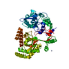 8azbC  4fajS S: Starting model for refinement C: citing same article ( |
|---|---|
| Similar structure data |
-Links
 PDBj
PDBj


- Assembly
| Deposited unit | 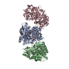
| ||||||||
|---|---|---|---|---|---|---|---|---|---|
| 1 | 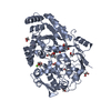
| ||||||||
| 2 | 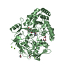
| ||||||||
| 3 | 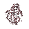
| ||||||||
| Unit cell |
|
-Components
| #1: Protein | Mass: 59501.270 Da / Num. of mol.: 3 Source method: isolated from a genetically manipulated source Source: (gene. exp.) #2: Chemical |   Mass: 390.389 Da / Num. of mol.: 3 / Source method: obtained synthetically / Formula: C15H26N4O8 / Feature type: SUBJECT OF INVESTIGATION Mass: 390.389 Da / Num. of mol.: 3 / Source method: obtained synthetically / Formula: C15H26N4O8 / Feature type: SUBJECT OF INVESTIGATION#3: Chemical | ChemComp-MG /   Mass: 24.305 Da / Num. of mol.: 4 / Source method: obtained synthetically / Formula: Mg Mass: 24.305 Da / Num. of mol.: 4 / Source method: obtained synthetically / Formula: Mg#4: Chemical | ChemComp-EDO /   Mass: 62.068 Da / Num. of mol.: 23 / Source method: obtained synthetically / Formula: C2H6O2 Mass: 62.068 Da / Num. of mol.: 23 / Source method: obtained synthetically / Formula: C2H6O2#5: Water | ChemComp-HOH / |  Mass: 18.015 Da / Num. of mol.: 1206 / Source method: isolated from a natural source / Formula: H2O Mass: 18.015 Da / Num. of mol.: 1206 / Source method: isolated from a natural source / Formula: H2OHas ligand of interest | Y | |
|---|
-Experimental details
-Experiment
| Experiment | Method: X-RAY DIFFRACTION / Number of used crystals: 1 |
|---|
- Sample preparation
| Crystal | Density Matthews: 2.23 Å3/Da / Density % sol: 44.86 % |
|---|---|
| Crystal grow | Temperature: 291 K / Method: vapor diffusion, hanging drop / pH: 8.5 Details: Crystals of DppE suitable for X-ray analysis were obtained from hanging drops formed by mixing 1 mircol of reservoir solution containing 0.1 M Bis-Tris-Propane pH 8.5, 0.4 M MgCl2, 22.5 % ...Details: Crystals of DppE suitable for X-ray analysis were obtained from hanging drops formed by mixing 1 mircol of reservoir solution containing 0.1 M Bis-Tris-Propane pH 8.5, 0.4 M MgCl2, 22.5 % PEG 3350 and 2.5 % DMSO with 1 microl of protein at 13 mg.ml-1 . |
-Data collection
| Diffraction | Mean temperature: 100 K / Serial crystal experiment: N | ||||||||||||||||||
|---|---|---|---|---|---|---|---|---|---|---|---|---|---|---|---|---|---|---|---|
| Diffraction source | Source: SYNCHROTRON / Site: Diamond / Beamline: I04 / Wavelength: 0.9795 Å | ||||||||||||||||||
| Detector | Type: DECTRIS PILATUS 6M / Detector: PIXEL / Date: Feb 14, 2015 | ||||||||||||||||||
| Radiation | Protocol: SINGLE WAVELENGTH / Monochromatic (M) / Laue (L): M / Scattering type: x-ray | ||||||||||||||||||
| Radiation wavelength | Wavelength: 0.9795 Å / Relative weight: 1 | ||||||||||||||||||
| Reflection | Resolution: 1.51→60.74 Å / Num. obs: 233391 / % possible obs: 96.3 % / Redundancy: 2.2 % / CC1/2: 0.998 / Rmerge(I) obs: 0.053 / Rpim(I) all: 0.053 / Rrim(I) all: 0.075 / Net I/σ(I): 8.4 | ||||||||||||||||||
| Reflection shell | Diffraction-ID: 1 / Redundancy: 2.2 %
|
- Processing
| Software |
| |||||||||||||||||||||||||||||||||||||||||||||||||||||||||||||||||||||||||||||||||||||||||||||||||||||||||||||||||||||||||||||||||||||||||||||||||||||||||||||||||||||||||||||||||||||||||||||||||||||||||||||||||||||||||||||||||||||||
|---|---|---|---|---|---|---|---|---|---|---|---|---|---|---|---|---|---|---|---|---|---|---|---|---|---|---|---|---|---|---|---|---|---|---|---|---|---|---|---|---|---|---|---|---|---|---|---|---|---|---|---|---|---|---|---|---|---|---|---|---|---|---|---|---|---|---|---|---|---|---|---|---|---|---|---|---|---|---|---|---|---|---|---|---|---|---|---|---|---|---|---|---|---|---|---|---|---|---|---|---|---|---|---|---|---|---|---|---|---|---|---|---|---|---|---|---|---|---|---|---|---|---|---|---|---|---|---|---|---|---|---|---|---|---|---|---|---|---|---|---|---|---|---|---|---|---|---|---|---|---|---|---|---|---|---|---|---|---|---|---|---|---|---|---|---|---|---|---|---|---|---|---|---|---|---|---|---|---|---|---|---|---|---|---|---|---|---|---|---|---|---|---|---|---|---|---|---|---|---|---|---|---|---|---|---|---|---|---|---|---|---|---|---|---|---|---|---|---|---|---|---|---|---|---|---|---|---|---|---|---|---|---|
| Refinement | Method to determine structure: MOLECULAR REPLACEMENT Starting model: 4FAJ Resolution: 1.51→60.739 Å / Cor.coef. Fo:Fc: 0.969 / Cor.coef. Fo:Fc free: 0.954 / SU B: 2.305 / SU ML: 0.079 / Cross valid method: FREE R-VALUE / ESU R: 0.081 / ESU R Free: 0.084 Details: Hydrogens have been added in their riding positions
| |||||||||||||||||||||||||||||||||||||||||||||||||||||||||||||||||||||||||||||||||||||||||||||||||||||||||||||||||||||||||||||||||||||||||||||||||||||||||||||||||||||||||||||||||||||||||||||||||||||||||||||||||||||||||||||||||||||||
| Solvent computation | Ion probe radii: 0.8 Å / Shrinkage radii: 0.8 Å / VDW probe radii: 1.2 Å / Solvent model: MASK BULK SOLVENT | |||||||||||||||||||||||||||||||||||||||||||||||||||||||||||||||||||||||||||||||||||||||||||||||||||||||||||||||||||||||||||||||||||||||||||||||||||||||||||||||||||||||||||||||||||||||||||||||||||||||||||||||||||||||||||||||||||||||
| Displacement parameters | Biso mean: 24.662 Å2
| |||||||||||||||||||||||||||||||||||||||||||||||||||||||||||||||||||||||||||||||||||||||||||||||||||||||||||||||||||||||||||||||||||||||||||||||||||||||||||||||||||||||||||||||||||||||||||||||||||||||||||||||||||||||||||||||||||||||
| Refinement step | Cycle: LAST / Resolution: 1.51→60.739 Å
| |||||||||||||||||||||||||||||||||||||||||||||||||||||||||||||||||||||||||||||||||||||||||||||||||||||||||||||||||||||||||||||||||||||||||||||||||||||||||||||||||||||||||||||||||||||||||||||||||||||||||||||||||||||||||||||||||||||||
| Refine LS restraints |
| |||||||||||||||||||||||||||||||||||||||||||||||||||||||||||||||||||||||||||||||||||||||||||||||||||||||||||||||||||||||||||||||||||||||||||||||||||||||||||||||||||||||||||||||||||||||||||||||||||||||||||||||||||||||||||||||||||||||
| LS refinement shell | Refine-ID: X-RAY DIFFRACTION / Total num. of bins used: 20
|
