 Movie
Movie Controller

[English] 日本語
 Yorodumi
- PDB-8awo: Crystal structure of a manganese-containing cupin (tm1459) from T... -
+ Open data

- Basic information
| Entry | Database: PDB / ID: 8awo | ||||||
|---|---|---|---|---|---|---|---|
| Title | Crystal structure of a manganese-containing cupin (tm1459) from Thermotoga maritima, variant AIFQ (Y7A/M38I/Y83F/C106Q) | ||||||
 Components Components | Cupin_2 domain-containing protein | ||||||
 Keywords Keywords | METAL BINDING PROTEIN / cupin / alkene cleavage / amino acid oxidation | ||||||
| Function / homology | Cupin 2, conserved barrel / Cupin domain / RmlC-like cupin domain superfamily / RmlC-like jelly roll fold / metal ion binding / Cupin type-2 domain-containing protein Function and homology information Function and homology information | ||||||
| Biological species |   Thermotoga maritima (bacteria) Thermotoga maritima (bacteria) | ||||||
| Method |  X-RAY DIFFRACTION / SYNCHROTRON / MOLECULAR REPLACEMENT / Resolution: 1.7 Å X-RAY DIFFRACTION / SYNCHROTRON / MOLECULAR REPLACEMENT / Resolution: 1.7 Å | ||||||
 Authors Authors | Grininger, C. / Steiner, K. / Gruber, K. / Pavkov-Keller, T. | ||||||
| Funding support |  Austria, 1items Austria, 1items
| ||||||
 Citation Citation | Journal: Chem Ing Tech / Year: 2023 Title: Engineering TM1459 for Stabilisation against Inactivation by Amino Acid Oxidation Authors: Grill, B. / Pavkov-Keller, T. / Grininger, C. / Darnhofer, B. / Gruber, K. / Hall, M. / Schwab, H. / Steiner, K. | ||||||
| History |
|
- Structure visualization
| Structure viewer | Molecule: MolmilJmol/JSmol |
|---|
- Downloads & links
-Download
| PDBx/mmCIF format | 8awo.cif.gz | 67.6 KB | Display | PDBx/mmCIF format |
|---|---|---|---|---|
| PDB format | pdb8awo.ent.gz | 44.8 KB | Display | PDB format |
| PDBx/mmJSON format | 8awo.json.gz | Tree view | PDBx/mmJSON format | |
| Others |  Other downloads Other downloads |
-Validation report
| Summary document | 8awo_validation.pdf.gz | 431.6 KB | Display | wwPDB validaton report |
|---|---|---|---|---|
| Full document | 8awo_full_validation.pdf.gz | 433.5 KB | Display | |
| Data in XML | 8awo_validation.xml.gz | 12.4 KB | Display | |
| Data in CIF | 8awo_validation.cif.gz | 17.3 KB | Display | |
| Arichive directory | https://data.pdbj.org/pub/pdb/validation_reports/aw/8awoftp://data.pdbj.org/pub/pdb/validation_reports/aw/8awo | HTTPS FTP |
-Related structure data
| Related structure data |  8awnC 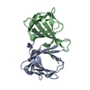 8awpC  1vj2S S: Starting model for refinement C: citing same article ( |
|---|---|
| Similar structure data |
-Links
 PDBj
PDBj- Assembly
| Deposited unit | 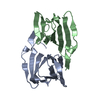
| ||||||||||||
|---|---|---|---|---|---|---|---|---|---|---|---|---|---|
| 1 |
| ||||||||||||
| Unit cell |
|
-Components
| #1: Protein | Mass: 13027.924 Da / Num. of mol.: 2 / Mutation: Y7A, M38I, Y83F, C106Q Source method: isolated from a genetically manipulated source Source: (gene. exp.) Thermotoga maritima (bacteria)Strain: ATCC 43589 / DSM 3109 / JCM 10099 / NBRC 100826 / MSB8 Gene: TM_1459 Production host: References: UniProt: Q9X1H0 #2: Water | ChemComp-HOH / |  Mass: 18.015 Da / Num. of mol.: 195 / Source method: isolated from a natural source / Formula: H2O Mass: 18.015 Da / Num. of mol.: 195 / Source method: isolated from a natural source / Formula: H2O |
|---|
-Experimental details
-Experiment
| Experiment | Method: X-RAY DIFFRACTION / Number of used crystals: 1 |
|---|
- Sample preparation
| Crystal | Density Matthews: 1.95 Å3/Da / Density % sol: 36.88 % |
|---|---|
| Crystal grow | Temperature: 289 K / Method: vapor diffusion, sitting drop / pH: 8.5 Details: 8 mg/ml protein concentration 0.1 M Tris pH 8.5 2 M ammonium sulfate |
-Data collection
| Diffraction | Mean temperature: 100 K / Serial crystal experiment: N |
|---|---|
| Diffraction source | Source: SYNCHROTRON / Site: ELETTRA  / Beamline: 5.2R / Wavelength: 1 Å / Beamline: 5.2R / Wavelength: 1 Å |
| Detector | Type: DECTRIS PILATUS 2M / Detector: PIXEL / Date: Mar 4, 2017 |
| Radiation | Protocol: SINGLE WAVELENGTH / Monochromatic (M) / Laue (L): M / Scattering type: x-ray |
| Radiation wavelength | Wavelength: 1 Å / Relative weight: 1 |
| Reflection | Resolution: 1.7→34.6 Å / Num. obs: 23041 / % possible obs: 99.75 % / Redundancy: 6.4 % / Biso Wilson estimate: 23.91 Å2 / CC1/2: 0.997 / CC star: 0.999 / Rmerge(I) obs: 0.08215 / Rpim(I) all: 0.03552 / Net I/σ(I): 12.75 |
| Reflection shell | Resolution: 1.7→1.761 Å / Rmerge(I) obs: 0.653 / Mean I/σ(I) obs: 2.82 / Num. unique obs: 2219 / CC1/2: 0.836 / CC star: 0.954 / Rpim(I) all: 0.2754 |
- Processing
| Software |
| |||||||||||||||||||||||||||||||||||||||||||||||||||||||||||||||
|---|---|---|---|---|---|---|---|---|---|---|---|---|---|---|---|---|---|---|---|---|---|---|---|---|---|---|---|---|---|---|---|---|---|---|---|---|---|---|---|---|---|---|---|---|---|---|---|---|---|---|---|---|---|---|---|---|---|---|---|---|---|---|---|---|
| Refinement | Method to determine structure: MOLECULAR REPLACEMENT Starting model: 1vj2 Resolution: 1.7→34.6 Å / SU ML: 0.2019 / Cross valid method: FREE R-VALUE / σ(F): 1.35 / Phase error: 24.1977 Stereochemistry target values: GeoStd + Monomer Library + CDL v1.2
| |||||||||||||||||||||||||||||||||||||||||||||||||||||||||||||||
| Solvent computation | Shrinkage radii: 0.9 Å / VDW probe radii: 1.11 Å / Solvent model: FLAT BULK SOLVENT MODEL | |||||||||||||||||||||||||||||||||||||||||||||||||||||||||||||||
| Displacement parameters | Biso mean: 28.96 Å2 | |||||||||||||||||||||||||||||||||||||||||||||||||||||||||||||||
| Refinement step | Cycle: LAST / Resolution: 1.7→34.6 Å
| |||||||||||||||||||||||||||||||||||||||||||||||||||||||||||||||
| Refine LS restraints |
| |||||||||||||||||||||||||||||||||||||||||||||||||||||||||||||||
| LS refinement shell |
|
