 Movie
Movie Controller

[English] 日本語
 Yorodumi
- PDB-8avu: Racemic protein crystal structure of aureocin A53 from Staphyloco... -
+ Open data

- Basic information
| Entry | Database: PDB / ID: 8avu | |||||||||||||||||||||||||||
|---|---|---|---|---|---|---|---|---|---|---|---|---|---|---|---|---|---|---|---|---|---|---|---|---|---|---|---|---|
| Title | Racemic protein crystal structure of aureocin A53 from Staphylococcus aureus in the dimeric state | |||||||||||||||||||||||||||
 Components Components |
| |||||||||||||||||||||||||||
 Keywords Keywords | ANTIMICROBIAL PROTEIN / Bacteriocin / D-protein / racemic / mirror-image | |||||||||||||||||||||||||||
| Function / homology | Bacteriocin class II, aureocin-like / Aureocin-like type II bacteriocin / killing of cells of another organism / defense response to bacterium / extracellular region / DI(HYDROXYETHYL)ETHER / polypeptide(D) / polypeptide(D) (> 10) / Bacteriocin aureocin A53 Function and homology information Function and homology information | |||||||||||||||||||||||||||
| Biological species |   Staphylococcus aureus (bacteria) Staphylococcus aureus (bacteria) | |||||||||||||||||||||||||||
| Method |  X-RAY DIFFRACTION / SYNCHROTRON / MOLECULAR REPLACEMENT / Resolution: 0.89 Å X-RAY DIFFRACTION / SYNCHROTRON / MOLECULAR REPLACEMENT / Resolution: 0.89 Å | |||||||||||||||||||||||||||
 Authors Authors | Lander, A.J. / Baumann, P. / Rizkallah, P. / Jin, Y. / Luk, L.Y.P. | |||||||||||||||||||||||||||
| Funding support |  United Kingdom, 8items United Kingdom, 8items
| |||||||||||||||||||||||||||
 Citation Citation | Journal: Commun Chem / Year: 2023 Title: Roles of inter- and intramolecular tryptophan interactions in membrane-active proteins revealed by racemic protein crystallography. Authors: Lander, A.J. / Mercado, L.D. / Li, X. / Taily, I.M. / Findlay, B.L. / Jin, Y. / Luk, L.Y.P. | |||||||||||||||||||||||||||
| History |
|
- Structure visualization
| Structure viewer | Molecule: MolmilJmol/JSmol |
|---|
- Downloads & links
-Download
| PDBx/mmCIF format | 8avu.cif.gz | 65.7 KB | Display | PDBx/mmCIF format |
|---|---|---|---|---|
| PDB format | pdb8avu.ent.gz | 46.8 KB | Display | PDB format |
| PDBx/mmJSON format | 8avu.json.gz | Tree view | PDBx/mmJSON format | |
| Others |  Other downloads Other downloads |
-Validation report
| Arichive directory | https://data.pdbj.org/pub/pdb/validation_reports/av/8avuftp://data.pdbj.org/pub/pdb/validation_reports/av/8avu | HTTPS FTP |
|---|
-Related structure data
| Related structure data |  7p5rC  8avrC  8avsC 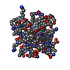 8avtC  2n8oS S: Starting model for refinement C: citing same article ( |
|---|---|
| Similar structure data |
-Links
 PDBj
PDBj- Assembly
| Deposited unit | 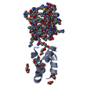
| ||||||||
|---|---|---|---|---|---|---|---|---|---|
| 1 | 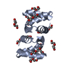
| ||||||||
| 2 | 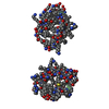
| ||||||||
| Unit cell |
|
-Components
| #1: Protein | Mass: 5994.228 Da / Num. of mol.: 1 / Source method: obtained synthetically / Source: (synth.) Staphylococcus aureus (bacteria) / References: UniProt: Q8GPI4 | ||||||||
|---|---|---|---|---|---|---|---|---|---|
| #2: Protein | Mass: 5984.158 Da / Num. of mol.: 1 / Source method: obtained synthetically / Source: (synth.) Staphylococcus aureus (bacteria) | ||||||||
| #3: Chemical |   Mass: 62.068 Da / Num. of mol.: 3 / Source method: obtained synthetically / Formula: C2H6O2 Mass: 62.068 Da / Num. of mol.: 3 / Source method: obtained synthetically / Formula: C2H6O2#4: Chemical | ChemComp-PEG /   Mass: 106.120 Da / Num. of mol.: 5 / Source method: obtained synthetically / Formula: C4H10O3 Mass: 106.120 Da / Num. of mol.: 5 / Source method: obtained synthetically / Formula: C4H10O3#5: Water | ChemComp-HOH / |  Mass: 18.015 Da / Num. of mol.: 143 / Source method: isolated from a natural source / Formula: H2O Mass: 18.015 Da / Num. of mol.: 143 / Source method: isolated from a natural source / Formula: H2OHas ligand of interest | N | Has protein modification | N | |
-Experimental details
-Experiment
| Experiment | Method: X-RAY DIFFRACTION / Number of used crystals: 1 |
|---|
- Sample preparation
| Crystal | Density Matthews: 1.84 Å3/Da / Density % sol: 33.03 % |
|---|---|
| Crystal grow | Temperature: 292 K / Method: vapor diffusion, sitting drop / pH: 8.5 Details: A racemic mixture of L-Aureocin A53 and D-Aureocin A53 at 40 mg/mL concentration was mixed 1:1 with 0.7 M sodium citrate, 0.1 M Tris precipitant condition, pH 8.5 in a 0.4 microlitre sitting ...Details: A racemic mixture of L-Aureocin A53 and D-Aureocin A53 at 40 mg/mL concentration was mixed 1:1 with 0.7 M sodium citrate, 0.1 M Tris precipitant condition, pH 8.5 in a 0.4 microlitre sitting drop. Crystals were submerged in 2.0 M lithium sulphate as cryoprotectant and flash frozen in liquid nitrogen during harvesting. |
-Data collection
| Diffraction | Mean temperature: 100 K / Serial crystal experiment: N |
|---|---|
| Diffraction source | Source: SYNCHROTRON / Site: Diamond / Beamline: I03 / Wavelength: 0.8153 Å |
| Detector | Type: DECTRIS EIGER2 XE 16M / Detector: PIXEL / Date: Nov 28, 2020 |
| Radiation | Protocol: SINGLE WAVELENGTH / Monochromatic (M) / Laue (L): M / Scattering type: x-ray |
| Radiation wavelength | Wavelength: 0.8153 Å / Relative weight: 1 |
| Reflection | Resolution: 0.89→21.99 Å / Num. obs: 62368 / % possible obs: 92.7 % / Redundancy: 5.9 % / CC1/2: 1 / Net I/σ(I): 17.2 |
| Reflection shell | Resolution: 0.89→0.91 Å / Redundancy: 3.5 % / Mean I/σ(I) obs: 1 / Num. unique obs: 1522 / CC1/2: 0.831 / % possible all: 46.4 |
- Processing
| Software |
| |||||||||||||||||||||||||||||||||||||||||||||||||||||||||||||||||||||||||||||||||||||||||||||||||||||||||||||||||||||||||||||||||||||||||||||||||||||||||||
|---|---|---|---|---|---|---|---|---|---|---|---|---|---|---|---|---|---|---|---|---|---|---|---|---|---|---|---|---|---|---|---|---|---|---|---|---|---|---|---|---|---|---|---|---|---|---|---|---|---|---|---|---|---|---|---|---|---|---|---|---|---|---|---|---|---|---|---|---|---|---|---|---|---|---|---|---|---|---|---|---|---|---|---|---|---|---|---|---|---|---|---|---|---|---|---|---|---|---|---|---|---|---|---|---|---|---|---|---|---|---|---|---|---|---|---|---|---|---|---|---|---|---|---|---|---|---|---|---|---|---|---|---|---|---|---|---|---|---|---|---|---|---|---|---|---|---|---|---|---|---|---|---|---|---|---|---|
| Refinement | Method to determine structure: MOLECULAR REPLACEMENT Starting model: 2N8O Resolution: 0.89→21.99 Å / Cor.coef. Fo:Fc: 0.974 / Cor.coef. Fo:Fc free: 0.968 / SU B: 0.78 / SU ML: 0.02 / Cross valid method: FREE R-VALUE / ESU R: 0.026 / ESU R Free: 0.026 Details: Hydrogens have been added in their riding positions
| |||||||||||||||||||||||||||||||||||||||||||||||||||||||||||||||||||||||||||||||||||||||||||||||||||||||||||||||||||||||||||||||||||||||||||||||||||||||||||
| Solvent computation | Ion probe radii: 0.8 Å / Shrinkage radii: 0.8 Å / VDW probe radii: 1.2 Å / Solvent model: MASK BULK SOLVENT | |||||||||||||||||||||||||||||||||||||||||||||||||||||||||||||||||||||||||||||||||||||||||||||||||||||||||||||||||||||||||||||||||||||||||||||||||||||||||||
| Displacement parameters | Biso mean: 11.304 Å2
| |||||||||||||||||||||||||||||||||||||||||||||||||||||||||||||||||||||||||||||||||||||||||||||||||||||||||||||||||||||||||||||||||||||||||||||||||||||||||||
| Refinement step | Cycle: LAST / Resolution: 0.89→21.99 Å
| |||||||||||||||||||||||||||||||||||||||||||||||||||||||||||||||||||||||||||||||||||||||||||||||||||||||||||||||||||||||||||||||||||||||||||||||||||||||||||
| Refine LS restraints |
| |||||||||||||||||||||||||||||||||||||||||||||||||||||||||||||||||||||||||||||||||||||||||||||||||||||||||||||||||||||||||||||||||||||||||||||||||||||||||||
| LS refinement shell |
|
