 Movie
Movie Controller


+ Open data

- Basic information
| Entry | Database: PDB / ID: 8asu | ||||||
|---|---|---|---|---|---|---|---|
| Title | The Crystal structure of short F46Y agroavidin - biotin complex | ||||||
 Components Components | Agroavidin | ||||||
 Keywords Keywords | UNKNOWN FUNCTION / BIOTIN BINDING PROTEIN / avidin / biotin / high affinity systems | ||||||
| Function / homology | Avidin/streptavidin / Avidin-like superfamily / Avidin family / Avidin-like domain profile. / biotin binding / extracellular region / BIOTIN / Avidin Function and homology information Function and homology information | ||||||
| Biological species |  Rhizobium sp. AAP43 (bacteria) Rhizobium sp. AAP43 (bacteria) | ||||||
| Method |  X-RAY DIFFRACTION / SYNCHROTRON / MOLECULAR REPLACEMENT / Resolution: 2.52 Å X-RAY DIFFRACTION / SYNCHROTRON / MOLECULAR REPLACEMENT / Resolution: 2.52 Å | ||||||
 Authors Authors | Livnah, O. / Bana, J. | ||||||
| Funding support |  Israel, 1items Israel, 1items
| ||||||
 Citation Citation | Journal: Febs J. / Year: 2023 Title: Self-assembly of a dimeric avidin into unique higher-order oligomers. Authors: Bana, J. / Warwar, J. / Bayer, E.A. / Livnah, O. | ||||||
| History |
|
- Structure visualization
| Structure viewer | Molecule: MolmilJmol/JSmol |
|---|
- Downloads & links
-Download
| PDBx/mmCIF format | 8asu.cif.gz | 102.2 KB | Display | PDBx/mmCIF format |
|---|---|---|---|---|
| PDB format | pdb8asu.ent.gz | 78 KB | Display | PDB format |
| PDBx/mmJSON format | 8asu.json.gz | Tree view | PDBx/mmJSON format | |
| Others |  Other downloads Other downloads |
-Validation report
| Summary document | 8asu_validation.pdf.gz | 1.3 MB | Display | wwPDB validaton report |
|---|---|---|---|---|
| Full document | 8asu_full_validation.pdf.gz | 1.4 MB | Display | |
| Data in XML | 8asu_validation.xml.gz | 19.5 KB | Display | |
| Data in CIF | 8asu_validation.cif.gz | 25.8 KB | Display | |
| Arichive directory | https://data.pdbj.org/pub/pdb/validation_reports/as/8asuftp://data.pdbj.org/pub/pdb/validation_reports/as/8asu | HTTPS FTP |
-Related structure data
| Related structure data |  8amhC  8an6C  8asrC  8assSC 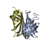 8astC 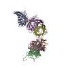 8avjC  8avpC C: citing same article ( S: Starting model for refinement |
|---|---|
| Similar structure data |

-Links
 PDBj
PDBj- Assembly
| Deposited unit | 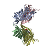
| ||||||||
|---|---|---|---|---|---|---|---|---|---|
| 1 | 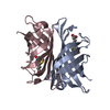
| ||||||||
| 2 | 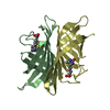
| ||||||||
| Unit cell |
|
-Components
| #1: Protein | Mass: 13794.177 Da / Num. of mol.: 4 / Mutation: F46Y Source method: isolated from a genetically manipulated source Source: (gene. exp.) Rhizobium sp. AAP43 (bacteria) / Gene: IP76_08565 / Production host: #2: Chemical | ChemComp-BTN /   Mass: 244.311 Da / Num. of mol.: 4 / Source method: obtained synthetically / Formula: C10H16N2O3S / Feature type: SUBJECT OF INVESTIGATION Mass: 244.311 Da / Num. of mol.: 4 / Source method: obtained synthetically / Formula: C10H16N2O3S / Feature type: SUBJECT OF INVESTIGATION#3: Water | ChemComp-HOH / |  Mass: 18.015 Da / Num. of mol.: 19 / Source method: isolated from a natural source / Formula: H2O Mass: 18.015 Da / Num. of mol.: 19 / Source method: isolated from a natural source / Formula: H2OHas ligand of interest | Y | Has protein modification | Y | |
|---|
-Experimental details
-Experiment
| Experiment | Method: X-RAY DIFFRACTION / Number of used crystals: 1 |
|---|
- Sample preparation
| Crystal | Density Matthews: 2.06 Å3/Da / Density % sol: 40.25 % |
|---|---|
| Crystal grow | Temperature: 293 K / Method: vapor diffusion, sitting drop / Details: 20% PEG 3350, 0.1 M Bis Tris |
-Data collection
| Diffraction | Mean temperature: 100 K / Serial crystal experiment: N |
|---|---|
| Diffraction source | Source: SYNCHROTRON / Site: ESRF  / Beamline: ID23-1 / Wavelength: 1.0723 Å / Beamline: ID23-1 / Wavelength: 1.0723 Å |
| Detector | Type: DECTRIS EIGER2 S 16M / Detector: PIXEL / Date: Nov 23, 2018 |
| Radiation | Protocol: SINGLE WAVELENGTH / Monochromatic (M) / Laue (L): M / Scattering type: x-ray |
| Radiation wavelength | Wavelength: 1.0723 Å / Relative weight: 1 |
| Reflection | Resolution: 2.52→57.75 Å / Num. obs: 15697 / % possible obs: 98.5 % / Redundancy: 4.6 % / CC1/2: 0.998 / Net I/σ(I): 11.2 |
| Reflection shell | Resolution: 2.52→2.59 Å / Redundancy: 4.7 % / Num. unique obs: 1118 / CC1/2: 0.507 / Rsym value: 0.923 / % possible all: 98.5 |
- Processing
| Software |
| ||||||||||||||||||||||||||||||||||||||||||||||||||||||||||||||||||||||||||||||||||||||||||||||||||||||||||||||||||||||||||||||||||||||||||||||||||||||||||||||||||||||||||||||||||||||
|---|---|---|---|---|---|---|---|---|---|---|---|---|---|---|---|---|---|---|---|---|---|---|---|---|---|---|---|---|---|---|---|---|---|---|---|---|---|---|---|---|---|---|---|---|---|---|---|---|---|---|---|---|---|---|---|---|---|---|---|---|---|---|---|---|---|---|---|---|---|---|---|---|---|---|---|---|---|---|---|---|---|---|---|---|---|---|---|---|---|---|---|---|---|---|---|---|---|---|---|---|---|---|---|---|---|---|---|---|---|---|---|---|---|---|---|---|---|---|---|---|---|---|---|---|---|---|---|---|---|---|---|---|---|---|---|---|---|---|---|---|---|---|---|---|---|---|---|---|---|---|---|---|---|---|---|---|---|---|---|---|---|---|---|---|---|---|---|---|---|---|---|---|---|---|---|---|---|---|---|---|---|---|---|
| Refinement | Method to determine structure: MOLECULAR REPLACEMENT Starting model: 8ASS Resolution: 2.52→44.09 Å / Cor.coef. Fo:Fc: 0.938 / Cor.coef. Fo:Fc free: 0.922 / SU B: 14.969 / SU ML: 0.317 / Cross valid method: THROUGHOUT / ESU R: 1.2 / ESU R Free: 0.344 / Stereochemistry target values: MAXIMUM LIKELIHOOD / Details: HYDROGENS HAVE BEEN ADDED IN THE RIDING POSITIONS
| ||||||||||||||||||||||||||||||||||||||||||||||||||||||||||||||||||||||||||||||||||||||||||||||||||||||||||||||||||||||||||||||||||||||||||||||||||||||||||||||||||||||||||||||||||||||
| Solvent computation | Ion probe radii: 0.8 Å / Shrinkage radii: 0.8 Å / VDW probe radii: 1.2 Å / Solvent model: MASK | ||||||||||||||||||||||||||||||||||||||||||||||||||||||||||||||||||||||||||||||||||||||||||||||||||||||||||||||||||||||||||||||||||||||||||||||||||||||||||||||||||||||||||||||||||||||
| Displacement parameters | Biso mean: 59.054 Å2
| ||||||||||||||||||||||||||||||||||||||||||||||||||||||||||||||||||||||||||||||||||||||||||||||||||||||||||||||||||||||||||||||||||||||||||||||||||||||||||||||||||||||||||||||||||||||
| Refinement step | Cycle: 1 / Resolution: 2.52→44.09 Å
| ||||||||||||||||||||||||||||||||||||||||||||||||||||||||||||||||||||||||||||||||||||||||||||||||||||||||||||||||||||||||||||||||||||||||||||||||||||||||||||||||||||||||||||||||||||||
| Refine LS restraints |
|