 Movie
Movie Controller


+ Open data

- Basic information
| Entry | Database: PDB / ID: 8ase | ||||||
|---|---|---|---|---|---|---|---|
| Title | Crystal structure of Thrombin in complex with macrocycle T3 | ||||||
 Components Components |
| ||||||
 Keywords Keywords | HYDROLASE / Serine endopeptidases / alpha-thrombin / activated factor IIa / fibrinogenase | ||||||
| Function / homology |  Function and homology information Function and homology informationcytolysis by host of symbiont cells / thrombospondin receptor activity / Defective factor XII causes hereditary angioedema / thrombin / thrombin-activated receptor signaling pathway / negative regulation of astrocyte differentiation / regulation of blood coagulation / positive regulation of phospholipase C-activating G protein-coupled receptor signaling pathway / neutrophil-mediated killing of gram-negative bacterium / Defective F8 cleavage by thrombin ...cytolysis by host of symbiont cells / thrombospondin receptor activity / Defective factor XII causes hereditary angioedema / thrombin / thrombin-activated receptor signaling pathway / negative regulation of astrocyte differentiation / regulation of blood coagulation / positive regulation of phospholipase C-activating G protein-coupled receptor signaling pathway / neutrophil-mediated killing of gram-negative bacterium / Defective F8 cleavage by thrombin / Platelet Aggregation (Plug Formation) / ligand-gated ion channel signaling pathway / positive regulation of collagen biosynthetic process / negative regulation of platelet activation / negative regulation of blood coagulation / positive regulation of blood coagulation / negative regulation of fibrinolysis / regulation of cytosolic calcium ion concentration / Transport of gamma-carboxylated protein precursors from the endoplasmic reticulum to the Golgi apparatus / Gamma-carboxylation of protein precursors / Common Pathway of Fibrin Clot Formation / Removal of aminoterminal propeptides from gamma-carboxylated proteins / fibrinolysis / Intrinsic Pathway of Fibrin Clot Formation / negative regulation of proteolysis / negative regulation of cytokine production involved in inflammatory response / Peptide ligand-binding receptors / Regulation of Complement cascade / positive regulation of release of sequestered calcium ion into cytosol / acute-phase response / Cell surface interactions at the vascular wall / positive regulation of receptor signaling pathway via JAK-STAT / growth factor activity / lipopolysaccharide binding / positive regulation of insulin secretion / platelet activation / response to wounding / positive regulation of protein localization to nucleus / Golgi lumen / Regulation of Insulin-like Growth Factor (IGF) transport and uptake by Insulin-like Growth Factor Binding Proteins (IGFBPs) / positive regulation of reactive oxygen species metabolic process / blood coagulation / antimicrobial humoral immune response mediated by antimicrobial peptide / regulation of cell shape / heparin binding / : / Thrombin signalling through proteinase activated receptors (PARs) / positive regulation of cell growth / blood microparticle / G alpha (q) signalling events / cell surface receptor signaling pathway / positive regulation of phosphatidylinositol 3-kinase/protein kinase B signal transduction / receptor ligand activity / endoplasmic reticulum lumen / signaling receptor binding / serine-type endopeptidase activity / positive regulation of cell population proliferation / calcium ion binding / proteolysis / extracellular space / extracellular exosome / extracellular region / plasma membrane Similarity search - Function | ||||||
| Biological species |  Homo sapiens (human) Homo sapiens (human) | ||||||
| Method |  X-RAY DIFFRACTION / SYNCHROTRON / MOLECULAR REPLACEMENT / Resolution: 2.55 Å X-RAY DIFFRACTION / SYNCHROTRON / MOLECULAR REPLACEMENT / Resolution: 2.55 Å | ||||||
 Authors Authors | Chinellato, M. / Angelini, A. / Nielsen, A. / Heinis, C. / Cendron, L. | ||||||
| Funding support |  Italy, 1items Italy, 1items
| ||||||
 Citation Citation | Journal: To Be Published Title: Crystal structure of Thrombin in complex with optimized macrocycles T1 and T3 Authors: Nielsen, A. / Chinellato, M. / Cendron, L. / Angelini, A. / Heinis, C. | ||||||
| History |
|
- Structure visualization
| Structure viewer | Molecule: MolmilJmol/JSmol |
|---|
- Downloads & links
-Download
| PDBx/mmCIF format | 8ase.cif.gz | 131.8 KB | Display | PDBx/mmCIF format |
|---|---|---|---|---|
| PDB format | pdb8ase.ent.gz | 100.6 KB | Display | PDB format |
| PDBx/mmJSON format | 8ase.json.gz | Tree view | PDBx/mmJSON format | |
| Others |  Other downloads Other downloads |
-Validation report
| Summary document | 8ase_validation.pdf.gz | 1.5 MB | Display | wwPDB validaton report |
|---|---|---|---|---|
| Full document | 8ase_full_validation.pdf.gz | 1.5 MB | Display | |
| Data in XML | 8ase_validation.xml.gz | 25.2 KB | Display | |
| Data in CIF | 8ase_validation.cif.gz | 33.6 KB | Display | |
| Arichive directory | https://data.pdbj.org/pub/pdb/validation_reports/as/8aseftp://data.pdbj.org/pub/pdb/validation_reports/as/8ase | HTTPS FTP |
-Related structure data
| Related structure data | 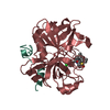 8asfC 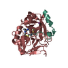 6z48S S: Starting model for refinement C: citing same article ( |
|---|---|
| Similar structure data |
-Links
 PDBj
PDBj


- Assembly
| Deposited unit | 
| ||||||||
|---|---|---|---|---|---|---|---|---|---|
| 1 | 
| ||||||||
| 2 | 
| ||||||||
| Unit cell |
|
-Components
-Protein/peptide / Protein , 2 types, 4 molecules LAHB
| #1: Protein/peptide | Mass: 4096.534 Da / Num. of mol.: 2 Source method: isolated from a genetically manipulated source Details: missing residues are not visible in the electron density map Source: (gene. exp.) Homo sapiens (human) / Gene: F2 / Production host: Homo sapiens (human) / References: UniProt: P00734, thrombin#2: Protein | Mass: 29780.219 Da / Num. of mol.: 2 Source method: isolated from a genetically manipulated source Details: missing residues are not visible in the electron density maps Source: (gene. exp.) Homo sapiens (human) / Gene: F2 / Production host: Homo sapiens (human) / References: UniProt: P00734, thrombin |
|---|
-Sugars , 2 types, 2 molecules 
| #3: Polysaccharide | 2-acetamido-2-deoxy-beta-D-glucopyranose-(1-4)-2-acetamido-2-deoxy-beta-D-glucopyranose |
|---|---|
| #6: Sugar | ChemComp-NAG /  Type: D-saccharide, beta linking / Mass: 221.208 Da / Num. of mol.: 1 / Source method: isolated from a natural source / Formula: C8H15NO6 Type: D-saccharide, beta linking / Mass: 221.208 Da / Num. of mol.: 1 / Source method: isolated from a natural source / Formula: C8H15NO6 |
-Non-polymers , 3 types, 81 molecules 


| #4: Chemical |  Mass: 586.208 Da / Num. of mol.: 2 / Source method: obtained synthetically / Formula: C30H36ClN3O3S2 Mass: 586.208 Da / Num. of mol.: 2 / Source method: obtained synthetically / Formula: C30H36ClN3O3S2#5: Chemical |  Mass: 96.063 Da / Num. of mol.: 2 / Source method: isolated from a natural source / Formula: SO4 Mass: 96.063 Da / Num. of mol.: 2 / Source method: isolated from a natural source / Formula: SO4#7: Water | ChemComp-HOH / | Mass: 18.015 Da / Num. of mol.: 77 / Source method: isolated from a natural source / Formula: H2O |
|---|
-Details
| Has ligand of interest | Y |
|---|---|
| Has protein modification | Y |
-Experimental details
-Experiment
| Experiment | Method: X-RAY DIFFRACTION / Number of used crystals: 1 |
|---|
- Sample preparation
| Crystal | Density Matthews: 2.65 Å3/Da / Density % sol: 53.52 % |
|---|---|
| Crystal grow | Temperature: 293 K / Method: vapor diffusion, hanging drop / pH: 5.5 Details: 100 mM Bis-Tris, 200 mM ammonium sulfate, 25% w/v PEG 3350 pH 5.5 |
-Data collection
| Diffraction | Mean temperature: 100 K / Serial crystal experiment: N |
|---|---|
| Diffraction source | Source: SYNCHROTRON / Site: ESRF  / Beamline: ID23-1 / Wavelength: 0.8856 Å / Beamline: ID23-1 / Wavelength: 0.8856 Å |
| Detector | Type: DECTRIS PILATUS 6M / Detector: PIXEL / Date: Jun 4, 2022 |
| Radiation | Protocol: SINGLE WAVELENGTH / Monochromatic (M) / Laue (L): M / Scattering type: x-ray |
| Radiation wavelength | Wavelength: 0.8856 Å / Relative weight: 1 |
| Reflection | Resolution: 2.55→41.81 Å / Num. obs: 23267 / % possible obs: 100 % / Redundancy: 9.7 % / CC1/2: 0.996 / Rmerge(I) obs: 0.18 / Net I/σ(I): 10 |
| Reflection shell | Resolution: 2.55→2.66 Å / Num. unique obs: 2943 / CC1/2: 0.77 |
- Processing
| Software |
| ||||||||||||||||||||
|---|---|---|---|---|---|---|---|---|---|---|---|---|---|---|---|---|---|---|---|---|---|
| Refinement | Method to determine structure: MOLECULAR REPLACEMENT Starting model: 6Z48 Resolution: 2.55→41.81 Å / Cross valid method: THROUGHOUT
| ||||||||||||||||||||
| Displacement parameters | Biso max: 127.59 Å2 / Biso mean: 45.5826 Å2 / Biso min: 18.43 Å2 | ||||||||||||||||||||
| Refinement step | Cycle: LAST / Resolution: 2.55→41.81 Å
|
