Mass: 18.015 Da / Num. of mol.: 1402 / Source method: isolated from a natural source / Formula: H2O
Has ligand of interest
Y
-
Experimental details
-
Experiment
Experiment
Method: X-RAY DIFFRACTION / Number of used crystals: 1
-
Sample preparation
Crystal
Density Matthews: 2.45 Å3/Da / Density % sol: 49.76 %
Crystal grow
Temperature: 292 K / Method: vapor diffusion, sitting drop / pH: 9.3 Details: 0.1 M BICINE pH 9.3, 22% v/v PEG Smear Board, 50 mM glycine, raltitrexed (powder was added); 1 uL of this solution supplemented with 50% ethylene glycol was added to the drop with crystals for cryoprotection
-
Data collection
Diffraction
Mean temperature: 100 K / Serial crystal experiment: N
Diffraction source
Source: SYNCHROTRON / Site: PETRA III, EMBL c/o DESY / Beamline: P13 (MX1) / Wavelength: 0.977 Å
Detector
Type: DECTRIS EIGER X 16M / Detector: PIXEL / Date: Nov 23, 2021
Radiation
Protocol: SINGLE WAVELENGTH / Monochromatic (M) / Laue (L): M / Scattering type: x-ray
Resolution: 1.23→55.15 Å / SU ML: 0.15 / Cross valid method: FREE R-VALUE / σ(F): 1.34 / Phase error: 36.5 / Stereochemistry target values: ML Details: Hydrogen atoms at riding positions were added for refinement. B factor of non H atoms were refined anisotropically
Rfactor
Num. reflection
% reflection
Selection details
Rfree
0.2294
1608
0.48 %
RANDOM
Rwork
0.1949
-
-
-
obs
0.195
332420
59.66 %
-
Solvent computation
Shrinkage radii: 0.9 Å / VDW probe radii: 1.1 Å / Solvent model: FLAT BULK SOLVENT MODEL
Refinement step
Cycle: LAST / Resolution: 1.23→55.15 Å
Protein
Nucleic acid
Ligand
Solvent
Total
Num. atoms
14213
0
112
1402
15727
Refine LS restraints
Refine-ID
Type
Dev ideal
X-RAY DIFFRACTION
f_bond_d
0.011
X-RAY DIFFRACTION
f_angle_d
1.134
X-RAY DIFFRACTION
f_dihedral_angle_d
13.847
X-RAY DIFFRACTION
f_chiral_restr
0.077
X-RAY DIFFRACTION
f_plane_restr
0.011
LS refinement shell
Resolution (Å)
Rfactor Rfree
Num. reflection Rfree
Rfactor Rwork
Num. reflection Rwork
Refine-ID
% reflection obs (%)
1.23-1.27
0.2581
18
0.2271
3819
X-RAY DIFFRACTION
8
1.27-1.32
0.2612
37
0.2224
7643
X-RAY DIFFRACTION
15
1.32-1.37
0.2755
55
0.2178
12627
X-RAY DIFFRACTION
25
1.37-1.43
0.2265
82
0.2302
16538
X-RAY DIFFRACTION
33
1.43-1.51
0.2442
99
0.2242
22176
X-RAY DIFFRACTION
44
1.51-1.6
0.2251
149
0.2194
30163
X-RAY DIFFRACTION
60
1.6-1.73
0.2382
177
0.2153
38551
X-RAY DIFFRACTION
76
1.73-1.9
0.2881
242
0.2107
47640
X-RAY DIFFRACTION
95
1.9-2.18
0.2289
245
0.1949
50417
X-RAY DIFFRACTION
100
2.18-2.74
0.2237
258
0.197
50440
X-RAY DIFFRACTION
100
2.74-55.15
0.2163
246
0.1808
50798
X-RAY DIFFRACTION
100
+
About Yorodumi
-
News
-
Feb 9, 2022. New format data for meta-information of EMDB entries
New format data for meta-information of EMDB entries
Version 3 of the EMDB header file is now the official format.
The previous official version 1.9 will be removed from the archive.
In the structure databanks used in Yorodumi, some data are registered as the other names, "COVID-19 virus" and "2019-nCoV". Here are the details of the virus and the list of structure data.
Jan 31, 2019. EMDB accession codes are about to change! (news from PDBe EMDB page)
EMDB accession codes are about to change! (news from PDBe EMDB page)
The allocation of 4 digits for EMDB accession codes will soon come to an end. Whilst these codes will remain in use, new EMDB accession codes will include an additional digit and will expand incrementally as the available range of codes is exhausted. The current 4-digit format prefixed with “EMD-” (i.e. EMD-XXXX) will advance to a 5-digit format (i.e. EMD-XXXXX), and so on. It is currently estimated that the 4-digit codes will be depleted around Spring 2019, at which point the 5-digit format will come into force.
The EM Navigator/Yorodumi systems omit the EMD- prefix.
Related info.:Q: What is EMD? / ID/Accession-code notation in Yorodumi/EM Navigator
Yorodumi is a browser for structure data from EMDB, PDB, SASBDB, etc.
This page is also the successor to EM Navigator detail page, and also detail information page/front-end page for Omokage search.
The word "yorodu" (or yorozu) is an old Japanese word meaning "ten thousand". "mi" (miru) is to see.
Related info.:EMDB / PDB / SASBDB / Comparison of 3 databanks / Yorodumi Search / Aug 31, 2016. New EM Navigator & Yorodumi / Yorodumi Papers / Jmol/JSmol / Function and homology information / Changes in new EM Navigator and Yorodumi
 Movie
Movie Controller


 Open data
 Basic information
 Components
 Keywords
 Function and homology information
 Homo sapiens (human)
 X-RAY DIFFRACTION /
  Authors
 Citation
 Structure visualization
 Downloads & links
 Other downloads
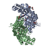
 PDBj
PDBj

 Assembly
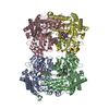


 Mass: 306.209 Da / Num. of mol.: 4 / Source method: obtained synthetically / Formula: C10H15N2O7P / Feature type: SUBJECT OF INVESTIGATION
 Mass: 62.068 Da / Num. of mol.: 8 / Source method: isolated from a natural source / Formula: C2H6O2
 Mass: 18.015 Da / Num. of mol.: 1402 / Source method: isolated from a natural source / Formula: H2O
 Sample preparation
 / Beamline: P13 (MX1) / Wavelength: 0.977 Å
 Processing