 Movie
Movie Controller

[English] 日本語
 Yorodumi
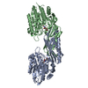
- PDB-8apz: Crystal structure of wild-type L-N-Carbamoylase from Sinorhizobiu... -
+ Open data

- Basic information
| Entry | Database: PDB / ID: 8apz | ||||||
|---|---|---|---|---|---|---|---|
| Title | Crystal structure of wild-type L-N-Carbamoylase from Sinorhizobium meliloti | ||||||
 Components Components | N-carbamoyl-L-amino-acid hydrolase | ||||||
 Keywords Keywords | HYDROLASE / peptidase family M20/M25/M40 | ||||||
| Function / homology |  Function and homology information Function and homology informationformylmethionine deformylase activity / N-carbamoyl-L-amino-acid hydrolase / N-carbamoyl-L-amino-acid hydrolase activity / hydrolase activity, acting on carbon-nitrogen (but not peptide) bonds, in linear amidines / amino acid biosynthetic process / metal ion binding Similarity search - Function | ||||||
| Biological species |  Sinorhizobium meliloti (bacteria) Sinorhizobium meliloti (bacteria) | ||||||
| Method |  X-RAY DIFFRACTION / SYNCHROTRON / MOLECULAR REPLACEMENT / molecular replacement / Resolution: 1.75 Å X-RAY DIFFRACTION / SYNCHROTRON / MOLECULAR REPLACEMENT / molecular replacement / Resolution: 1.75 Å | ||||||
 Authors Authors | Rozeboom, H.J. / Mayer, C. | ||||||
| Funding support |  Netherlands, 1items Netherlands, 1items
| ||||||
 Citation Citation | Journal: Angew.Chem.Int.Ed.Engl. / Year: 2023 Title: Selecting Better Biocatalysts by Complementing Recoded Bacteria. Authors: Rubini, R. / Jansen, S.C. / Beekhuis, H. / Rozeboom, H.J. / Mayer, C. | ||||||
| History |
|
- Structure visualization
| Structure viewer | Molecule: MolmilJmol/JSmol |
|---|
- Downloads & links
-Download
| PDBx/mmCIF format | 8apz.cif.gz | 326.8 KB | Display | PDBx/mmCIF format |
|---|---|---|---|---|
| PDB format | pdb8apz.ent.gz | 261.8 KB | Display | PDB format |
| PDBx/mmJSON format | 8apz.json.gz | Tree view | PDBx/mmJSON format | |
| Others |  Other downloads Other downloads |
-Validation report
| Arichive directory | https://data.pdbj.org/pub/pdb/validation_reports/ap/8apzftp://data.pdbj.org/pub/pdb/validation_reports/ap/8apz | HTTPS FTP |
|---|
-Related structure data
-Links
 PDBj
PDBj
- Assembly
| Deposited unit | 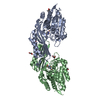
| ||||||||
|---|---|---|---|---|---|---|---|---|---|
| 1 |
| ||||||||
| Unit cell |
|
-Components
-Protein , 1 types, 2 molecules AB
| #1: Protein | Mass: 47039.250 Da / Num. of mol.: 2 Source method: isolated from a genetically manipulated source Details: First 21 residues are expression tag / Source: (gene. exp.) Sinorhizobium meliloti (bacteria) / Gene: hyuC / Plasmid: pACYCDUET-1 / Production host: References: UniProt: Q6DTN4, N-carbamoyl-L-amino-acid hydrolase |
|---|
-Non-polymers , 6 types, 476 molecules 

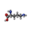


| #2: Chemical | ChemComp-ZN /  Mass: 65.409 Da / Num. of mol.: 4 / Source method: obtained synthetically / Formula: Zn Mass: 65.409 Da / Num. of mol.: 4 / Source method: obtained synthetically / Formula: Zn#3: Chemical | ChemComp-FE /  Mass: 55.845 Da / Num. of mol.: 4 / Source method: obtained synthetically / Formula: Fe Mass: 55.845 Da / Num. of mol.: 4 / Source method: obtained synthetically / Formula: Fe#4: Chemical |  Type: D-peptide linking / Mass: 132.161 Da / Num. of mol.: 2 / Source method: obtained synthetically / Formula: C5H12N2O2 Type: D-peptide linking / Mass: 132.161 Da / Num. of mol.: 2 / Source method: obtained synthetically / Formula: C5H12N2O2#5: Chemical |  Mass: 59.044 Da / Num. of mol.: 3 / Source method: obtained synthetically / Formula: C2H3O2 Mass: 59.044 Da / Num. of mol.: 3 / Source method: obtained synthetically / Formula: C2H3O2#6: Chemical |  Mass: 92.094 Da / Num. of mol.: 3 / Source method: obtained synthetically / Formula: C3H8O3 Mass: 92.094 Da / Num. of mol.: 3 / Source method: obtained synthetically / Formula: C3H8O3#7: Water | ChemComp-HOH / | Mass: 18.015 Da / Num. of mol.: 460 / Source method: isolated from a natural source / Formula: H2O |
|---|
-Details
| Has ligand of interest | N |
|---|
-Experimental details
-Experiment
| Experiment | Method: X-RAY DIFFRACTION / Number of used crystals: 1 |
|---|
- Sample preparation
| Crystal | Density Matthews: 2 Å3/Da / Density % sol: 38 % |
|---|---|
| Crystal grow | Temperature: 294 K / Method: vapor diffusion, sitting drop / pH: 6.5 Details: 0.2 M sodium acetate, 0.1 M Bis-tris propane and 20 % PEG3350 |
-Data collection
| Diffraction | Mean temperature: 100 K / Serial crystal experiment: N |
|---|---|
| Diffraction source | Source: SYNCHROTRON / Site: ESRF  / Beamline: MASSIF-1 / Wavelength: 0.9655 Å / Beamline: MASSIF-1 / Wavelength: 0.9655 Å |
| Detector | Type: DECTRIS PILATUS 2M / Detector: PIXEL / Date: Feb 22, 2022 |
| Radiation | Protocol: SINGLE WAVELENGTH / Monochromatic (M) / Laue (L): M / Scattering type: x-ray |
| Radiation wavelength | Wavelength: 0.9655 Å / Relative weight: 1 |
| Reflection | Resolution: 1.75→45.53 Å / Num. obs: 74615 / % possible obs: 99.5 % / Redundancy: 6.5 % / CC1/2: 0.997 / Rmerge(I) obs: 0.088 / Rpim(I) all: 0.037 / Rrim(I) all: 0.095 / Net I/σ(I): 13.3 / Num. measured all: 482305 |
| Reflection shell | Resolution: 1.75→1.78 Å / Redundancy: 6.2 % / Rmerge(I) obs: 0.778 / Num. unique obs: 3970 / CC1/2: 0.836 / Rpim(I) all: 0.332 / Rrim(I) all: 0.848 / % possible all: 97.4 |
-Phasing
| Phasing | Method: molecular replacement | |||||||||
|---|---|---|---|---|---|---|---|---|---|---|
| Phasing MR | Model details: Phaser MODE: MR_AUTO
|
- Processing
| Software |
| |||||||||||||||||||||||||||||||||||||||||||||||||||||||||||||||||||||||||||
|---|---|---|---|---|---|---|---|---|---|---|---|---|---|---|---|---|---|---|---|---|---|---|---|---|---|---|---|---|---|---|---|---|---|---|---|---|---|---|---|---|---|---|---|---|---|---|---|---|---|---|---|---|---|---|---|---|---|---|---|---|---|---|---|---|---|---|---|---|---|---|---|---|---|---|---|---|
| Refinement | Method to determine structure: MOLECULAR REPLACEMENT Starting model: AF model Resolution: 1.75→43.22 Å / Cor.coef. Fo:Fc: 0.974 / Cor.coef. Fo:Fc free: 0.959 / SU B: 4.916 / SU ML: 0.077 / Cross valid method: THROUGHOUT / σ(F): 0 / ESU R: 0.104 / ESU R Free: 0.102 / Stereochemistry target values: MAXIMUM LIKELIHOOD Details: U VALUES : WITH TLS ADDED HYDROGENS HAVE BEEN ADDED IN THE RIDING POSITIONS
| |||||||||||||||||||||||||||||||||||||||||||||||||||||||||||||||||||||||||||
| Solvent computation | Ion probe radii: 0.7 Å / Shrinkage radii: 0.7 Å / VDW probe radii: 1.4 Å / Solvent model: MASK | |||||||||||||||||||||||||||||||||||||||||||||||||||||||||||||||||||||||||||
| Displacement parameters | Biso max: 83.18 Å2 / Biso mean: 25.558 Å2 / Biso min: 13.88 Å2
| |||||||||||||||||||||||||||||||||||||||||||||||||||||||||||||||||||||||||||
| Refinement step | Cycle: final / Resolution: 1.75→43.22 Å
| |||||||||||||||||||||||||||||||||||||||||||||||||||||||||||||||||||||||||||
| Refine LS restraints |
| |||||||||||||||||||||||||||||||||||||||||||||||||||||||||||||||||||||||||||
| LS refinement shell | Resolution: 1.75→1.795 Å / Rfactor Rfree error: 0 / Total num. of bins used: 20
| |||||||||||||||||||||||||||||||||||||||||||||||||||||||||||||||||||||||||||
| Refinement TLS params. | Method: refined / Refine-ID: X-RAY DIFFRACTION
| |||||||||||||||||||||||||||||||||||||||||||||||||||||||||||||||||||||||||||
| Refinement TLS group |
|
