 Movie
Movie Controller


+ Open data

- Basic information
| Entry | Database: PDB / ID: 8anu | ||||||
|---|---|---|---|---|---|---|---|
| Title | Crystal structure of protein phi3T-93 | ||||||
 Components Components | YopN. Phi3T_93 | ||||||
 Keywords Keywords | VIRAL PROTEIN / Arbitrium / Phi3T / lysis-lysogeny decission | ||||||
| Function / homology | metal ion binding / NICKEL (II) ION / Uncharacterized protein Function and homology information Function and homology information | ||||||
| Biological species |  Bacillus phage phi3T (virus) Bacillus phage phi3T (virus) | ||||||
| Method |  X-RAY DIFFRACTION / SYNCHROTRON / MOLECULAR REPLACEMENT / Resolution: 2.31014705408 Å X-RAY DIFFRACTION / SYNCHROTRON / MOLECULAR REPLACEMENT / Resolution: 2.31014705408 Å | ||||||
 Authors Authors | Zamora-Caballero, S. / Marina, A. | ||||||
| Funding support |  Spain, 1items Spain, 1items
| ||||||
 Citation Citation | Journal: Nat Microbiol / Year: 2024 Title: Antagonistic interactions between phage and host factors control arbitrium lysis-lysogeny decision. Authors: Zamora-Caballero, S. / Chmielowska, C. / Quiles-Puchalt, N. / Brady, A. / Del Sol, F.G. / Mancheno-Bonillo, J. / Felipe-Ruiz, A. / Meijer, W.J.J. / Penades, J.R. / Marina, A. | ||||||
| History |
|
- Structure visualization
| Structure viewer | Molecule: MolmilJmol/JSmol |
|---|
- Downloads & links
-Download
| PDBx/mmCIF format | 8anu.cif.gz | 78.2 KB | Display | PDBx/mmCIF format |
|---|---|---|---|---|
| PDB format | pdb8anu.ent.gz | 51.4 KB | Display | PDB format |
| PDBx/mmJSON format | 8anu.json.gz | Tree view | PDBx/mmJSON format | |
| Others |  Other downloads Other downloads |
-Validation report
| Summary document | 8anu_validation.pdf.gz | 1.4 MB | Display | wwPDB validaton report |
|---|---|---|---|---|
| Full document | 8anu_full_validation.pdf.gz | 1.4 MB | Display | |
| Data in XML | 8anu_validation.xml.gz | 7.3 KB | Display | |
| Data in CIF | 8anu_validation.cif.gz | 9.2 KB | Display | |
| Arichive directory | https://data.pdbj.org/pub/pdb/validation_reports/an/8anuftp://data.pdbj.org/pub/pdb/validation_reports/an/8anu | HTTPS FTP |
-Related structure data
| Related structure data | 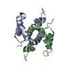 8antSC 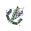 8anvC 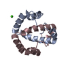 8c8eC S: Starting model for refinement C: citing same article ( |
|---|---|
| Similar structure data |
-Links
 PDBj
PDBj- Assembly
| Deposited unit | 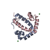
| |||||||||||||||||||||||||||||||||||||||||||||||||||||||||||||||||||||||||||||||||||||||||||||||||||||||||
|---|---|---|---|---|---|---|---|---|---|---|---|---|---|---|---|---|---|---|---|---|---|---|---|---|---|---|---|---|---|---|---|---|---|---|---|---|---|---|---|---|---|---|---|---|---|---|---|---|---|---|---|---|---|---|---|---|---|---|---|---|---|---|---|---|---|---|---|---|---|---|---|---|---|---|---|---|---|---|---|---|---|---|---|---|---|---|---|---|---|---|---|---|---|---|---|---|---|---|---|---|---|---|---|---|---|---|
| 1 |
| |||||||||||||||||||||||||||||||||||||||||||||||||||||||||||||||||||||||||||||||||||||||||||||||||||||||||
| Unit cell |
| |||||||||||||||||||||||||||||||||||||||||||||||||||||||||||||||||||||||||||||||||||||||||||||||||||||||||
| Components on special symmetry positions |
| |||||||||||||||||||||||||||||||||||||||||||||||||||||||||||||||||||||||||||||||||||||||||||||||||||||||||
| Noncrystallographic symmetry (NCS) | NCS domain:
NCS domain segments: Ens-ID: 1
|
-Components
| #1: Protein | Mass: 9768.052 Da / Num. of mol.: 2 Source method: isolated from a genetically manipulated source Source: (gene. exp.) Bacillus phage phi3T (virus) / Gene: phi3T_93 / Production host:  #2: Chemical |   Mass: 58.693 Da / Num. of mol.: 2 / Source method: obtained synthetically / Formula: Ni / Feature type: SUBJECT OF INVESTIGATION Mass: 58.693 Da / Num. of mol.: 2 / Source method: obtained synthetically / Formula: Ni / Feature type: SUBJECT OF INVESTIGATION#3: Water | ChemComp-HOH / |  Mass: 18.015 Da / Num. of mol.: 36 / Source method: isolated from a natural source / Formula: H2O Mass: 18.015 Da / Num. of mol.: 36 / Source method: isolated from a natural source / Formula: H2OHas ligand of interest | Y | |
|---|
-Experimental details
-Experiment
| Experiment | Method: X-RAY DIFFRACTION / Number of used crystals: 1 |
|---|
- Sample preparation
| Crystal | Density Matthews: 1.99 Å3/Da / Density % sol: 38.15 % |
|---|---|
| Crystal grow | Temperature: 294.15 K / Method: vapor diffusion, sitting drop Details: 10%PEG8000, 50mM magnesium acetate, 100mM sodium acetate |
-Data collection
| Diffraction | Mean temperature: 100 K / Serial crystal experiment: N |
|---|---|
| Diffraction source | Source: SYNCHROTRON / Site: ALBA / Beamline: XALOC / Wavelength: 0.97918 Å |
| Detector | Type: DECTRIS PILATUS 6M / Detector: PIXEL / Date: Oct 28, 2021 |
| Radiation | Monochromator: Channel-cut Si (111) / Protocol: SINGLE WAVELENGTH / Monochromatic (M) / Laue (L): M / Scattering type: x-ray |
| Radiation wavelength | Wavelength: 0.97918 Å / Relative weight: 1 |
| Reflection | Resolution: 2.31→43.47 Å / Num. obs: 6697 / % possible obs: 98.1 % / Redundancy: 4.9 % / Biso Wilson estimate: 33.4114979572 Å2 / CC1/2: 0.993 / Rmerge(I) obs: 0.1 / Net I/σ(I): 8.5 |
| Reflection shell | Resolution: 2.31→2.44 Å / Redundancy: 5.1 % / Rmerge(I) obs: 0.212 / Mean I/σ(I) obs: 4.4 / Num. unique obs: 985 / CC1/2: 0.961 / Rpim(I) all: 0.102 / Rrim(I) all: 0.237 / % possible all: 98.6 |
- Processing
| Software |
| ||||||||||||||||||||||||
|---|---|---|---|---|---|---|---|---|---|---|---|---|---|---|---|---|---|---|---|---|---|---|---|---|---|
| Refinement | Method to determine structure: MOLECULAR REPLACEMENT Starting model: 8ANT Resolution: 2.31014705408→40.06302883 Å / SU ML: 0.252471672262 / Cross valid method: FREE R-VALUE / σ(F): 1.33786559852 / Phase error: 28.1250642639
| ||||||||||||||||||||||||
| Solvent computation | Shrinkage radii: 0.9 Å / VDW probe radii: 1.11 Å / Solvent model: FLAT BULK SOLVENT MODEL | ||||||||||||||||||||||||
| Displacement parameters | Biso mean: 38.1353823492 Å2 | ||||||||||||||||||||||||
| Refinement step | Cycle: LAST / Resolution: 2.31014705408→40.06302883 Å
| ||||||||||||||||||||||||
| Refine LS restraints |
| ||||||||||||||||||||||||
| LS refinement shell | Resolution: 2.31015→2.9104 Å
|
