 Movie
Movie Controller

[English] 日本語
 Yorodumi
- PDB-8an9: Fucosylated mixed-chirality linear peptide FHP5 bound to the fuco... -
+ Open data

- Basic information
| Entry | Database: PDB / ID: 8an9 | |||||||||
|---|---|---|---|---|---|---|---|---|---|---|
| Title | Fucosylated mixed-chirality linear peptide FHP5 bound to the fucose binding lectin LecB PA-IIL from Pseudomonas aeruginosa at 1.3 Angstrom resolution. | |||||||||
 Components Components |
| |||||||||
 Keywords Keywords | ANTIBIOTIC / Antimicrobial peptide / alpha helix / mixed-chirality | |||||||||
| Function / homology | Lectin, sugar-binding / Calcium-mediated lectin / Calcium-mediated lectin superfamily / Fucose-binding lectin II (PA-IIL) / single-species biofilm formation / carbohydrate binding / metal ion binding / Chem-ZDC / Fucose-binding lectin PA-IIL Function and homology information Function and homology information | |||||||||
| Biological species |  Pseudomonas aeruginosa PAO1 (bacteria) Pseudomonas aeruginosa PAO1 (bacteria)synthetic construct (others) | |||||||||
| Method |  X-RAY DIFFRACTION / SYNCHROTRON / MOLECULAR REPLACEMENT / molecular replacement / Resolution: 1.273 Å X-RAY DIFFRACTION / SYNCHROTRON / MOLECULAR REPLACEMENT / molecular replacement / Resolution: 1.273 Å | |||||||||
 Authors Authors | Personne, H. / Stocker, A. / Reymond, J.-L. | |||||||||
| Funding support | European Union,  Switzerland, 2items Switzerland, 2items
| |||||||||
 Citation Citation | Journal: J.Med.Chem. / Year: 2023 Title: To Fold or Not to Fold: Diastereomeric Optimization of an alpha-Helical Antimicrobial Peptide. Authors: Personne, H. / Paschoud, T. / Fulgencio, S. / Baeriswyl, S. / Kohler, T. / van Delden, C. / Stocker, A. / Javor, S. / Reymond, J.L. | |||||||||
| History |
|
- Structure visualization
| Structure viewer | Molecule: MolmilJmol/JSmol |
|---|
- Downloads & links
-Download
| PDBx/mmCIF format | 8an9.cif.gz | 200.7 KB | Display | PDBx/mmCIF format |
|---|---|---|---|---|
| PDB format | pdb8an9.ent.gz | Display | PDB format | |
| PDBx/mmJSON format | 8an9.json.gz | Tree view | PDBx/mmJSON format | |
| Others |  Other downloads Other downloads |
-Validation report
| Arichive directory | https://data.pdbj.org/pub/pdb/validation_reports/an/8an9ftp://data.pdbj.org/pub/pdb/validation_reports/an/8an9 | HTTPS FTP |
|---|
-Related structure data
| Related structure data |  8anoC  8anrC  8aooC 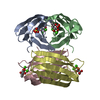 1oxcS S: Starting model for refinement C: citing same article ( |
|---|---|
| Similar structure data |
-Links
 PDBj
PDBj- Assembly
| Deposited unit | 
| ||||||||
|---|---|---|---|---|---|---|---|---|---|
| 1 |
| ||||||||
| Unit cell |
|
-Components
| #1: Protein | Mass: 11734.707 Da / Num. of mol.: 4 Source method: isolated from a genetically manipulated source Source: (gene. exp.) Pseudomonas aeruginosa PAO1 (bacteria) / Gene: lecB, PA3361 / Production host: #2: Protein/peptide | Mass: 1323.840 Da / Num. of mol.: 3 / Source method: obtained synthetically / Source: (synth.) synthetic construct (others) #3: Chemical | ChemComp-CA /   Mass: 40.078 Da / Num. of mol.: 8 / Source method: obtained synthetically / Formula: Ca Mass: 40.078 Da / Num. of mol.: 8 / Source method: obtained synthetically / Formula: Ca#4: Sugar | ChemComp-ZDC /   Type: D-saccharide / Mass: 206.193 Da / Num. of mol.: 4 / Source method: obtained synthetically / Formula: C8H14O6 / Feature type: SUBJECT OF INVESTIGATION Type: D-saccharide / Mass: 206.193 Da / Num. of mol.: 4 / Source method: obtained synthetically / Formula: C8H14O6 / Feature type: SUBJECT OF INVESTIGATION#5: Water | ChemComp-HOH / |  Mass: 18.015 Da / Num. of mol.: 482 / Source method: isolated from a natural source / Formula: H2O Mass: 18.015 Da / Num. of mol.: 482 / Source method: isolated from a natural source / Formula: H2OHas ligand of interest | Y | Has protein modification | Y | |
|---|
-Experimental details
-Experiment
| Experiment | Method: X-RAY DIFFRACTION / Number of used crystals: 1 |
|---|
- Sample preparation
| Crystal | Density Matthews: 2.13 Å3/Da / Density % sol: 42.16 % |
|---|---|
| Crystal grow | Temperature: 291.15 K / Method: vapor diffusion, sitting drop / pH: 7.5 / Details: 0.1 M HEPES pH 7.5, 20% Jeffamine M-600 |
-Data collection
| Diffraction | Mean temperature: 100 K / Serial crystal experiment: N |
|---|---|
| Diffraction source | Source: SYNCHROTRON / Site: SLS / Beamline: X06DA / Wavelength: 1.000023 Å |
| Detector | Type: DECTRIS PILATUS 2M-F / Detector: PIXEL / Date: Dec 13, 2021 |
| Radiation | Protocol: SINGLE WAVELENGTH / Monochromatic (M) / Laue (L): M / Scattering type: x-ray |
| Radiation wavelength | Wavelength: 1.000023 Å / Relative weight: 1 |
| Reflection | Resolution: 1.273→44.87 Å / Num. obs: 211407 / % possible obs: 96.9 % / Redundancy: 1.03 % / CC1/2: 0.998 / Rrim(I) all: 0.069 / Net I/σ(I): 9.16 |
| Reflection shell | Resolution: 1.273→1.279 Å / Redundancy: 2.4 % / Mean I/σ(I) obs: 1.08 / Num. unique obs: 32349 / CC1/2: 0.717 / Rrim(I) all: 0.841 / % possible all: 41.7 |
-Phasing
| Phasing | Method: molecular replacement |
|---|
- Processing
| Software |
| ||||||||||||||||||||||||||||||||||||||||||||||||||||||||||||||||||||||||||||||||||||||||||||||||||||||||||||||||||||||||||||||||||||||||||||||||||||||||||||||||||||||||||||||||||||||||||
|---|---|---|---|---|---|---|---|---|---|---|---|---|---|---|---|---|---|---|---|---|---|---|---|---|---|---|---|---|---|---|---|---|---|---|---|---|---|---|---|---|---|---|---|---|---|---|---|---|---|---|---|---|---|---|---|---|---|---|---|---|---|---|---|---|---|---|---|---|---|---|---|---|---|---|---|---|---|---|---|---|---|---|---|---|---|---|---|---|---|---|---|---|---|---|---|---|---|---|---|---|---|---|---|---|---|---|---|---|---|---|---|---|---|---|---|---|---|---|---|---|---|---|---|---|---|---|---|---|---|---|---|---|---|---|---|---|---|---|---|---|---|---|---|---|---|---|---|---|---|---|---|---|---|---|---|---|---|---|---|---|---|---|---|---|---|---|---|---|---|---|---|---|---|---|---|---|---|---|---|---|---|---|---|---|---|---|---|
| Refinement | Method to determine structure: MOLECULAR REPLACEMENT Starting model: 1OXC Resolution: 1.273→44.87 Å / SU ML: 0.13 / Cross valid method: THROUGHOUT / σ(F): 1.34 / Phase error: 20.81 / Stereochemistry target values: ML
| ||||||||||||||||||||||||||||||||||||||||||||||||||||||||||||||||||||||||||||||||||||||||||||||||||||||||||||||||||||||||||||||||||||||||||||||||||||||||||||||||||||||||||||||||||||||||||
| Solvent computation | Shrinkage radii: 0.9 Å / VDW probe radii: 1.11 Å / Solvent model: FLAT BULK SOLVENT MODEL | ||||||||||||||||||||||||||||||||||||||||||||||||||||||||||||||||||||||||||||||||||||||||||||||||||||||||||||||||||||||||||||||||||||||||||||||||||||||||||||||||||||||||||||||||||||||||||
| Displacement parameters | Biso max: 99.32 Å2 / Biso mean: 23.2965 Å2 / Biso min: 11.41 Å2 | ||||||||||||||||||||||||||||||||||||||||||||||||||||||||||||||||||||||||||||||||||||||||||||||||||||||||||||||||||||||||||||||||||||||||||||||||||||||||||||||||||||||||||||||||||||||||||
| Refinement step | Cycle: final / Resolution: 1.273→44.87 Å
| ||||||||||||||||||||||||||||||||||||||||||||||||||||||||||||||||||||||||||||||||||||||||||||||||||||||||||||||||||||||||||||||||||||||||||||||||||||||||||||||||||||||||||||||||||||||||||
| LS refinement shell | Refine-ID: X-RAY DIFFRACTION / Rfactor Rfree error: 0
|
