 Movie
Movie Controller

[English] 日本語
 Yorodumi
- PDB-8ai8: Crystal structure of glutathione transferase Chi 1 from Synechocy... -
+ Open data

- Basic information
| Entry | Database: PDB / ID: 8ai8 | ||||||
|---|---|---|---|---|---|---|---|
| Title | Crystal structure of glutathione transferase Chi 1 from Synechocystis sp. PCC 6803 in complex with glutathione | ||||||
 Components Components | Glutathione S-transferase family protein | ||||||
 Keywords Keywords | TRANSFERASE / Glutathione transferase / cyanobateria / chi class / glutathione | ||||||
| Function / homology |  Function and homology information Function and homology informationGlutathione S-transferase Yfyf (Class Pi); Chain A, domain 2 - #10 / Glutathione S-transferase Yfyf (Class Pi); Chain A, domain 2 / Glutaredoxin / Glutaredoxin / Up-down Bundle / 3-Layer(aba) Sandwich / Mainly Alpha / Alpha Beta Similarity search - Domain/homology | ||||||
| Biological species |  | ||||||
| Method |  X-RAY DIFFRACTION / SYNCHROTRON / MOLECULAR REPLACEMENT / Resolution: 1.7 Å X-RAY DIFFRACTION / SYNCHROTRON / MOLECULAR REPLACEMENT / Resolution: 1.7 Å | ||||||
 Authors Authors | Didierjean, C. | ||||||
| Funding support |  France, 1items France, 1items
| ||||||
 Citation Citation | Journal: Biomolecules / Year: 2022 Title: Biochemical and Structural Characterization of Chi-Class Glutathione Transferases: A Snapshot on the Glutathione Transferase Encoded by sll0067 Gene in the Cyanobacterium Synechocystis sp. Strain PCC 6803. Authors: Mocchetti, E. / Morette, L. / Mulliert, G. / Mathiot, S. / Guillot, B. / Dehez, F. / Chauvat, F. / Cassier-Chauvat, C. / Brochier-Armanet, C. / Didierjean, C. / Hecker, A. | ||||||
| History |
|
- Structure visualization
| Structure viewer | Molecule: MolmilJmol/JSmol |
|---|
- Downloads & links
-Download
| PDBx/mmCIF format | 8ai8.cif.gz | 97.8 KB | Display | PDBx/mmCIF format |
|---|---|---|---|---|
| PDB format | pdb8ai8.ent.gz | 74.2 KB | Display | PDB format |
| PDBx/mmJSON format | 8ai8.json.gz | Tree view | PDBx/mmJSON format | |
| Others |  Other downloads Other downloads |
-Validation report
| Arichive directory | https://data.pdbj.org/pub/pdb/validation_reports/ai/8ai8ftp://data.pdbj.org/pub/pdb/validation_reports/ai/8ai8 | HTTPS FTP |
|---|
-Related structure data
| Related structure data |  8ai9C  8aibC  3lszS S: Starting model for refinement C: citing same article ( |
|---|---|
| Similar structure data |
-Links
 PDBj
PDBj

- Assembly
| Deposited unit | 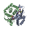
| ||||||||
|---|---|---|---|---|---|---|---|---|---|
| 1 |
| ||||||||
| Unit cell |
| ||||||||
| Components on special symmetry positions |
|
-Components
| #1: Protein | Mass: 21466.732 Da / Num. of mol.: 2 Source method: isolated from a genetically manipulated source Source: (gene. exp.) #2: Chemical |   Mass: 307.323 Da / Num. of mol.: 2 / Source method: obtained synthetically / Formula: C10H17N3O6S Mass: 307.323 Da / Num. of mol.: 2 / Source method: obtained synthetically / Formula: C10H17N3O6S#3: Water | ChemComp-HOH / |  Mass: 18.015 Da / Num. of mol.: 445 / Source method: isolated from a natural source / Formula: H2O Mass: 18.015 Da / Num. of mol.: 445 / Source method: isolated from a natural source / Formula: H2O |
|---|
-Experimental details
-Experiment
| Experiment | Method: X-RAY DIFFRACTION / Number of used crystals: 1 |
|---|
- Sample preparation
| Crystal grow | Temperature: 277 K / Method: microbatch Details: 16% (w/v) PEG 8000, 40 mM potassium phosphate monobasic and 20% (w/v) glycerol |
|---|
-Data collection
| Diffraction | Mean temperature: 100 K / Serial crystal experiment: N |
|---|---|
| Diffraction source | Source: SYNCHROTRON / Site: ESRF / Beamline: BM07 / Wavelength: 0.97951 Å |
| Detector | Type: DECTRIS PILATUS 6M / Detector: PIXEL / Date: Oct 11, 2021 |
| Radiation | Protocol: SINGLE WAVELENGTH / Monochromatic (M) / Laue (L): M / Scattering type: x-ray |
| Radiation wavelength | Wavelength: 0.97951 Å / Relative weight: 1 |
| Reflection | Resolution: 1.7→48.4 Å / Num. obs: 92986 / % possible obs: 100 % / Redundancy: 12.9 % / Biso Wilson estimate: 29.6 Å2 / CC1/2: 1 / Rmerge(I) obs: 0.058 / Net I/σ(I): 24.8 |
| Reflection shell | Resolution: 1.7→1.73 Å / Rmerge(I) obs: 1.168 / Mean I/σ(I) obs: 1.8 / Num. unique obs: 4529 / CC1/2: 0.832 |
- Processing
| Software |
| ||||||||||||||||||||||||||||||||||||||||||||||||||||||||||||||||||
|---|---|---|---|---|---|---|---|---|---|---|---|---|---|---|---|---|---|---|---|---|---|---|---|---|---|---|---|---|---|---|---|---|---|---|---|---|---|---|---|---|---|---|---|---|---|---|---|---|---|---|---|---|---|---|---|---|---|---|---|---|---|---|---|---|---|---|---|
| Refinement | Method to determine structure: MOLECULAR REPLACEMENT Starting model: 3LSZ Resolution: 1.7→24.8 Å / Cor.coef. Fo:Fc: 0.938 / Cor.coef. Fo:Fc free: 0.936 / SU R Cruickshank DPI: 0.073 / Cross valid method: THROUGHOUT / SU R Blow DPI: 0.078 / SU Rfree Blow DPI: 0.077 / SU Rfree Cruickshank DPI: 0.073
| ||||||||||||||||||||||||||||||||||||||||||||||||||||||||||||||||||
| Displacement parameters | Biso mean: 34.58 Å2
| ||||||||||||||||||||||||||||||||||||||||||||||||||||||||||||||||||
| Refine analyze | Luzzati coordinate error obs: 0.24 Å | ||||||||||||||||||||||||||||||||||||||||||||||||||||||||||||||||||
| Refinement step | Cycle: LAST / Resolution: 1.7→24.8 Å
| ||||||||||||||||||||||||||||||||||||||||||||||||||||||||||||||||||
| Refine LS restraints |
| ||||||||||||||||||||||||||||||||||||||||||||||||||||||||||||||||||
| LS refinement shell | Resolution: 1.7→1.71 Å
|
