 Movie
Movie Controller

[English] 日本語
 Yorodumi
- PDB-8ah5: Crystal Structure of the third PDZ domain of PSD-95 protein in th... -
+ Open data

- Basic information
| Entry | Database: PDB / ID: 8ah5 | ||||||
|---|---|---|---|---|---|---|---|
| Title | Crystal Structure of the third PDZ domain of PSD-95 protein in the space group P212121 at pH 4.6 | ||||||
 Components Components | cDNA FLJ50577, highly similar to Discs large homolog 4 | ||||||
 Keywords Keywords | PROTEIN BINDING / PDZ domain | ||||||
| Function / homology |  Function and homology information Function and homology informationneuromuscular junction / kinase binding / chemical synaptic transmission / neuron projection Similarity search - Function | ||||||
| Biological species |  Homo sapiens (human) Homo sapiens (human) | ||||||
| Method |  X-RAY DIFFRACTION / SYNCHROTRON / MOLECULAR REPLACEMENT / molecular replacement / Resolution: 1.25 Å X-RAY DIFFRACTION / SYNCHROTRON / MOLECULAR REPLACEMENT / molecular replacement / Resolution: 1.25 Å | ||||||
| Model details | pH 4.0 P3112 polymorph | ||||||
 Authors Authors | Camara-Artigas, A. / Salinas Garcia, M.C. | ||||||
| Funding support |  Spain, 1items Spain, 1items
| ||||||
 Citation Citation | Journal: Crystals / Year: 2023 Title: pH-Driven Polymorphic Behaviour of the Third PDZ Domain of PSD95: The Role of Electrostatic Interactions Authors: Salinas-Garcia, M.C. / Plaza-Garrido, M. / Gavira, J.A. / Murciano-Calles, J. / Andujar-Sanchez, M. / Ortiz-Salmeron, E. / Martinez, J.C. / Camara-Artigas, A. | ||||||
| History |
|
- Structure visualization
| Structure viewer | Molecule: MolmilJmol/JSmol |
|---|
- Downloads & links
-Download
| PDBx/mmCIF format | 8ah5.cif.gz | 75.1 KB | Display | PDBx/mmCIF format |
|---|---|---|---|---|
| PDB format | pdb8ah5.ent.gz | 55.7 KB | Display | PDB format |
| PDBx/mmJSON format | 8ah5.json.gz | Tree view | PDBx/mmJSON format | |
| Others |  Other downloads Other downloads |
-Validation report
| Summary document | 8ah5_validation.pdf.gz | 424 KB | Display | wwPDB validaton report |
|---|---|---|---|---|
| Full document | 8ah5_full_validation.pdf.gz | 424 KB | Display | |
| Data in XML | 8ah5_validation.xml.gz | 8 KB | Display | |
| Data in CIF | 8ah5_validation.cif.gz | 11.2 KB | Display | |
| Arichive directory | https://data.pdbj.org/pub/pdb/validation_reports/ah/8ah5ftp://data.pdbj.org/pub/pdb/validation_reports/ah/8ah5 | HTTPS FTP |
-Related structure data
| Related structure data | 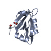 8ah4C  8ah6C  8ah7C  8ah8C 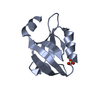 6qjiS S: Starting model for refinement C: citing same article ( |
|---|---|
| Similar structure data |
-Links
 PDBj
PDBj- Assembly
| Deposited unit | 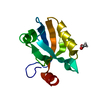
| ||||||||
|---|---|---|---|---|---|---|---|---|---|
| 1 |
| ||||||||
| Unit cell |
|
-Components
| #1: Protein | Mass: 11148.488 Da / Num. of mol.: 1 Source method: isolated from a genetically manipulated source Source: (gene. exp.) Homo sapiens (human) / Plasmid: pET15 / Production host:  | ||||
|---|---|---|---|---|---|
| #2: Chemical |   Mass: 59.044 Da / Num. of mol.: 2 / Source method: obtained synthetically / Formula: C2H3O2 Mass: 59.044 Da / Num. of mol.: 2 / Source method: obtained synthetically / Formula: C2H3O2#3: Water | ChemComp-HOH / |  Mass: 18.015 Da / Num. of mol.: 171 / Source method: isolated from a natural source / Formula: H2O Mass: 18.015 Da / Num. of mol.: 171 / Source method: isolated from a natural source / Formula: H2OHas ligand of interest | N | |
-Experimental details
-Experiment
| Experiment | Method: X-RAY DIFFRACTION / Number of used crystals: 1 |
|---|
- Sample preparation
| Crystal | Density Matthews: 1.85 Å3/Da / Density % sol: 33.42 % |
|---|---|
| Crystal grow | Temperature: 298 K / Method: vapor diffusion, sitting drop / pH: 4.6 / Details: 30% PEG4K, 0.2M AMSO4, 0.1M AcONa |
-Data collection
| Diffraction | Mean temperature: 100 K / Serial crystal experiment: N | ||||||||||||||||||||||||||||||
|---|---|---|---|---|---|---|---|---|---|---|---|---|---|---|---|---|---|---|---|---|---|---|---|---|---|---|---|---|---|---|---|
| Diffraction source | Source: SYNCHROTRON / Site: ESRF  / Beamline: ID30B / Wavelength: 0.9677 Å / Beamline: ID30B / Wavelength: 0.9677 Å | ||||||||||||||||||||||||||||||
| Detector | Type: DECTRIS EIGER X 4M / Detector: PIXEL / Date: Nov 18, 2021 | ||||||||||||||||||||||||||||||
| Radiation | Protocol: SINGLE WAVELENGTH / Monochromatic (M) / Laue (L): M / Scattering type: x-ray | ||||||||||||||||||||||||||||||
| Radiation wavelength | Wavelength: 0.9677 Å / Relative weight: 1 | ||||||||||||||||||||||||||||||
| Reflection | Resolution: 1.25→19.35 Å / Num. obs: 22582 / % possible obs: 95.6 % / Redundancy: 10.7 % / Biso Wilson estimate: 10.7 Å2 / CC1/2: 0.996 / Rmerge(I) obs: 0.13 / Rpim(I) all: 0.04 / Rrim(I) all: 0.136 / Net I/σ(I): 9.9 / Num. measured all: 242181 / Scaling rejects: 9 | ||||||||||||||||||||||||||||||
| Reflection shell | Diffraction-ID: 1
|
-Phasing
| Phasing | Method: molecular replacement |
|---|
- Processing
| Software |
| |||||||||||||||||||||||||||||||||||||||||||||||||||||||||||||||
|---|---|---|---|---|---|---|---|---|---|---|---|---|---|---|---|---|---|---|---|---|---|---|---|---|---|---|---|---|---|---|---|---|---|---|---|---|---|---|---|---|---|---|---|---|---|---|---|---|---|---|---|---|---|---|---|---|---|---|---|---|---|---|---|---|
| Refinement | Method to determine structure: MOLECULAR REPLACEMENT Starting model: 6QJI Resolution: 1.25→19.35 Å / SU ML: 0.12 / Cross valid method: THROUGHOUT / σ(F): 1 / Phase error: 21.3 / Stereochemistry target values: ML
| |||||||||||||||||||||||||||||||||||||||||||||||||||||||||||||||
| Solvent computation | Shrinkage radii: 0.9 Å / VDW probe radii: 1.1 Å / Solvent model: FLAT BULK SOLVENT MODEL | |||||||||||||||||||||||||||||||||||||||||||||||||||||||||||||||
| Displacement parameters | Biso max: 44.86 Å2 / Biso mean: 14.3678 Å2 / Biso min: 6.02 Å2 | |||||||||||||||||||||||||||||||||||||||||||||||||||||||||||||||
| Refinement step | Cycle: final / Resolution: 1.25→19.35 Å
| |||||||||||||||||||||||||||||||||||||||||||||||||||||||||||||||
| LS refinement shell | Refine-ID: X-RAY DIFFRACTION / Rfactor Rfree error: 0 / Total num. of bins used: 8
|
