regulation of mRNA stability involved in cellular response to UV / regulation of eIF2 alpha phosphorylation by dsRNA / regulation of endoribonuclease activity / negative regulation of centrosome duplication / positive regulation of cell cycle G2/M phase transition / regulation of endodeoxyribonuclease activity / regulation of centriole replication / granular component / positive regulation of centrosome duplication / positive regulation of protein localization to nucleolus ...regulation of mRNA stability involved in cellular response to UV / regulation of eIF2 alpha phosphorylation by dsRNA / regulation of endoribonuclease activity / negative regulation of centrosome duplication / positive regulation of cell cycle G2/M phase transition / regulation of endodeoxyribonuclease activity / regulation of centriole replication / granular component / positive regulation of centrosome duplication / positive regulation of protein localization to nucleolus / negative regulation of protein kinase activity by regulation of protein phosphorylation / TFAP2A acts as a transcriptional repressor during retinoic acid induced cell differentiation / synaptic target recognition / Golgi reassembly / SARS-CoV-1-host interactions / regulation of centrosome duplication / NOTCH4 Activation and Transmission of Signal to the Nucleus / establishment of Golgi localization / respiratory system process / spindle pole centrosome / ALK mutants bind TKIs / tube formation / Tat protein binding / regulation of synapse maturation / Rap1 signalling / Nuclear import of Rev protein / cell volume homeostasis / negative regulation of protein localization to nucleus / centrosome cycle / TP53 regulates transcription of additional cell cycle genes whose exact role in the p53 pathway remain uncertain / regulation of DNA damage response, signal transduction by p53 class mediator / nucleocytoplasmic transport / KSRP (KHSRP) binds and destabilizes mRNA / GP1b-IX-V activation signalling / protein kinase inhibitor activity / ribosomal large subunit binding / negative regulation of mRNA splicing, via spliceosome / macrophage differentiation / ribosomal small subunit binding / Interleukin-3, Interleukin-5 and GM-CSF signaling / Regulation of localization of FOXO transcription factors / ribosomal large subunit export from nucleus / phosphoserine residue binding / Activation of BAD and translocation to mitochondria / NF-kappaB binding / regulation of ERK1 and ERK2 cascade / protein targeting / Chk1/Chk2(Cds1) mediated inactivation of Cyclin B:Cdk1 complex / SARS-CoV-2 targets host intracellular signalling and regulatory pathways / core promoter sequence-specific DNA binding / cellular response to glucose starvation / Nuclear events stimulated by ALK signaling in cancer / SARS-CoV-1 targets host intracellular signalling and regulatory pathways / RHO GTPases activate PKNs / ribosomal small subunit export from nucleus / negative regulation of TORC1 signaling / ERK1 and ERK2 cascade / Transcriptional and post-translational regulation of MITF-M expression and activity / Deposition of new CENPA-containing nucleosomes at the centromere / lung development / SUMOylation of transcription cofactors / protein sequestering activity / negative regulation of innate immune response / positive regulation of translation / hippocampal mossy fiber to CA3 synapse / ribosomal large subunit biogenesis / positive regulation of protein ubiquitination / ribosome assembly / TP53 Regulates Metabolic Genes / Translocation of SLC2A4 (GLUT4) to the plasma membrane / Deactivation of the beta-catenin transactivating complex / intracellular protein transport / regulation of cell growth / Negative regulation of NOTCH4 signaling / positive regulation of NF-kappaB transcription factor activity / protein-DNA complex / PKR-mediated signaling / regulation of protein stability / protein import into nucleus / cellular response to UV / cellular senescence / Signaling by ALK fusions and activated point mutants / intracellular protein localization / melanosome / unfolded protein binding / nucleosome assembly / large ribosomal subunit / ribosomal small subunit biogenesis / small ribosomal subunit / angiogenesis / protein phosphatase binding / histone binding / blood microparticle / molecular adaptor activity / vesicle / DNA-binding transcription factor binding / transmembrane transporter binding / transcription coactivator activity / protein phosphorylation / rRNA binding Similarity search - Function
Resolution: 2.9→21.91 Å / Cor.coef. Fo:Fc: 0.929 / Cor.coef. Fo:Fc free: 0.887 / SU B: 64.541 / SU ML: 0.478 / SU R Cruickshank DPI: 2.3403 / Cross valid method: THROUGHOUT / σ(F): 0 / ESU R: 2.34 / ESU R Free: 0.479 / Stereochemistry target values: MAXIMUM LIKELIHOOD Details: HYDROGENS HAVE BEEN ADDED IN THE RIDING POSITIONS U VALUES : WITH TLS ADDED
Rfactor
Num. reflection
% reflection
Selection details
Rfree
0.3226
730
4.8 %
RANDOM
Rwork
0.2697
-
-
-
obs
0.2722
14615
93.84 %
-
Solvent computation
Ion probe radii: 0.8 Å / Shrinkage radii: 0.8 Å / VDW probe radii: 1.2 Å / Solvent model: MASK
In the structure databanks used in Yorodumi, some data are registered as the other names, "COVID-19 virus" and "2019-nCoV". Here are the details of the virus and the list of structure data.
Jan 31, 2019. EMDB accession codes are about to change! (news from PDBe EMDB page)
EMDB accession codes are about to change! (news from PDBe EMDB page)
The allocation of 4 digits for EMDB accession codes will soon come to an end. Whilst these codes will remain in use, new EMDB accession codes will include an additional digit and will expand incrementally as the available range of codes is exhausted. The current 4-digit format prefixed with “EMD-” (i.e. EMD-XXXX) will advance to a 5-digit format (i.e. EMD-XXXXX), and so on. It is currently estimated that the 4-digit codes will be depleted around Spring 2019, at which point the 5-digit format will come into force.
The EM Navigator/Yorodumi systems omit the EMD- prefix.
Related info.:Q: What is EMD? / ID/Accession-code notation in Yorodumi/EM Navigator
Yorodumi is a browser for structure data from EMDB, PDB, SASBDB, etc.
This page is also the successor to EM Navigator detail page, and also detail information page/front-end page for Omokage search.
The word "yorodu" (or yorozu) is an old Japanese word meaning "ten thousand". "mi" (miru) is to see.
Related info.:EMDB / PDB / SASBDB / Comparison of 3 databanks / Yorodumi Search / Aug 31, 2016. New EM Navigator & Yorodumi / Yorodumi Papers / Jmol/JSmol / Function and homology information / Changes in new EM Navigator and Yorodumi
 Movie
Movie Controller

 Yorodumi
 Open data
 Basic information
 Components
 Keywords
 Function and homology information
 Homo sapiens (human)
 X-RAY DIFFRACTION /
  Authors
 Russian Federation, 2items
  Citation
 Structure visualization
 Downloads & links
 Other downloads
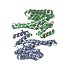
 PDBj
PDBj


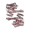
 Assembly


 Mass: 18.015 Da / Num. of mol.: 14 / Source method: isolated from a natural source / Formula: H2O
 Sample preparation
 Processing