[English] 日本語
Yorodumi
- PDB-8afg: K352D oxalyl-CoA synthetase Pcs60p -

+
Open data


ID or keywords:

Loading...

-
Basic information

Entry
Database: PDB / ID: 8afg
TitleK352D oxalyl-CoA synthetase Pcs60p
ComponentsOxalate--CoA ligase
KeywordsLIGASE / Peroxisome / Oxalyl-CoA ligase / Oligomer / Yeast
Function / homology
Function and homology information


oxalate-CoA ligase / oxalate-CoA ligase activity / medium-chain fatty acid-CoA ligase activity / oxalate catabolic process / peroxisomal membrane / peroxisomal matrix / fatty acid metabolic process / mRNA binding / ATP binding / cytoplasm
Similarity search - Function
Oxalate--CoA ligase Pcs60-like / ANL, N-terminal domain / AMP-binding enzyme, C-terminal domain / AMP-binding enzyme C-terminal domain / AMP-binding, conserved site / Putative AMP-binding domain signature. / AMP-dependent synthetase/ligase / AMP-binding enzyme, C-terminal domain superfamily / AMP-binding enzyme
Similarity search - Domain/homology
Biological speciesSaccharomyces cerevisiae (brewer's yeast)
MethodX-RAY DIFFRACTION / SYNCHROTRON / MOLECULAR REPLACEMENT / Resolution: 2.45 Å
AuthorsBurgi, J. / Chojnowski, G. / Wilmanns, M.
Funding support Germany, 3items
OrganizationGrant numberCountry
German Research Foundation (DFG)1058/9-1 Germany
German Research Foundation (DFG)1058/9-2 Germany
EIPOD fellowship under Marie Sklodowska-Curie Actions COFUND664726 Germany
CitationJournal: Biol Chem / Year: 2023
Title: Asymmetric horseshoe-like assembly of peroxisomal yeast oxalyl-CoA synthetase.
Authors: Jérôme Bürgi / Pascal Lill / Evdokia-Anastasia Giannopoulou / Cy M Jeffries / Grzegorz Chojnowski / Stefan Raunser / Christos Gatsogiannis / Matthias Wilmanns /
Abstract: Oxalyl-CoA synthetase from is one of the most abundant peroxisomal proteins in yeast and hence has become a model to study peroxisomal translocation. It contains a C-terminal Peroxisome Targeting ...Oxalyl-CoA synthetase from is one of the most abundant peroxisomal proteins in yeast and hence has become a model to study peroxisomal translocation. It contains a C-terminal Peroxisome Targeting Signal 1, which however is partly dispensable, suggesting additional receptor bindings sites. To unravel any additional features that may contribute to its capacity to be recognized as peroxisomal target, we determined its assembly and overall architecture by an integrated structural biology approach, including X-ray crystallography, single particle cryo-electron microscopy and small angle X-ray scattering. Surprisingly, it assembles into mixture of concentration-dependent dimers, tetramers and hexamers by dimer self-association. Hexameric particles form an unprecedented asymmetric horseshoe-like arrangement, which considerably differs from symmetric hexameric assembly found in many other protein structures. A single mutation within the self-association interface is sufficient to abolish any higher-level oligomerization, resulting in a homogenous dimeric assembly. The small C-terminal domain of yeast Oxalyl-CoA synthetase is connected by a partly flexible hinge with the large N-terminal domain, which provides the sole basis for oligomeric assembly. Our data provide a basis to mechanistically study peroxisomal translocation of this target.
History
DepositionJul 17, 2022Deposition site: PDBE / Processing site: PDBE
Revision 1.0Feb 8, 2023Provider: repository / Type: Initial release
Revision 1.1Feb 22, 2023Group: Database references / Category: citation / citation_author
Item: _citation.journal_volume / _citation.page_first ..._citation.journal_volume / _citation.page_first / _citation.page_last / _citation_author.identifier_ORCID
Revision 1.2May 1, 2024Group: Data collection / Refinement description
Category: chem_comp_atom / chem_comp_bond ...chem_comp_atom / chem_comp_bond / pdbx_initial_refinement_model / struct_ncs_dom_lim
Item: _struct_ncs_dom_lim.beg_auth_comp_id / _struct_ncs_dom_lim.beg_label_asym_id ..._struct_ncs_dom_lim.beg_auth_comp_id / _struct_ncs_dom_lim.beg_label_asym_id / _struct_ncs_dom_lim.beg_label_comp_id / _struct_ncs_dom_lim.beg_label_seq_id / _struct_ncs_dom_lim.end_auth_comp_id / _struct_ncs_dom_lim.end_label_asym_id / _struct_ncs_dom_lim.end_label_comp_id / _struct_ncs_dom_lim.end_label_seq_id

-
Structure visualization

Structure viewerMolecule:
MolmilJmol/JSmol

Downloads & links

-
Assembly

Deposited unit
A: Oxalate--CoA ligase
B: Oxalate--CoA ligase
hetero molecules


Theoretical massNumber of molelcules
Total (without water)121,6057
Polymers121,1252
Non-polymers4805
Water2,738152
1


  • Idetical with deposited unit
  • defined by author
  • Evidence: light scattering, SAXS, Stable dimer across all protein concentration tested.
TypeNameSymmetry operationNumber
identity operation1_555x,y,z1
Unit cell
Length a, b, c (Å)59.931, 125.600, 80.495
Angle α, β, γ (deg.)90.000, 96.929, 90.000
Int Tables number4
Space group name H-MP1211
Noncrystallographic symmetry (NCS)NCS domain:
IDEns-IDDetails
11A
21B

NCS domain segments:

Ens-ID: 1 / Beg auth comp-ID: ALA / Beg label comp-ID: ALA / End auth comp-ID: ILE / End label comp-ID: ILE / Auth seq-ID: 9 - 437 / Label seq-ID: 9 - 437

Dom-IDComponent-IDAuth asym-IDLabel asym-ID
11AA
22BB

NCS ensembles : (Details: Local NCS retraints between domains: 1 2)

-
Components

#1: Protein Oxalate--CoA ligase / Acyl-activating enzyme 3 / Oxalyl-CoA synthetase / Peroxisomal-coenzyme A synthetase


Mass: 60562.410 Da / Num. of mol.: 2
Source method: isolated from a genetically manipulated source
Source: (gene. exp.) Saccharomyces cerevisiae (brewer's yeast)
Strain: ATCC 204508 / S288c / Gene: PCS60, AAE3, FAT2, YBR222C, YBR1512 / Production host: Escherichia coli (E. coli) / References: UniProt: P38137, oxalate-CoA ligase
#2: Chemical
ChemComp-SO4 / SULFATE ION


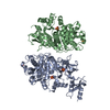
Mass: 96.063 Da / Num. of mol.: 5 / Source method: obtained synthetically / Formula: SO4
#3: Water ChemComp-HOH / water


Mass: 18.015 Da / Num. of mol.: 152 / Source method: isolated from a natural source / Formula: H2O
Has ligand of interestN

-
Experimental details

-
Experiment

ExperimentMethod: X-RAY DIFFRACTION / Number of used crystals: 1

-
Sample preparation

CrystalDensity Matthews: 2.49 Å3/Da / Density % sol: 50.57 %
Crystal growTemperature: 298 K / Method: vapor diffusion, sitting drop / pH: 6
Details: Ammonium sulfate 0.15M, MES 0.1M pH6, PEG 4000 15%(w/v)
PH range: 6

-
Data collection

DiffractionMean temperature: 100 K / Serial crystal experiment: N
Diffraction sourceSource: SYNCHROTRON / Site: PETRA III, EMBL c/o DESY / Beamline: P13 (MX1) / Wavelength: 0.987 Å
DetectorType: DECTRIS PILATUS3 6M / Detector: PIXEL / Date: Jun 5, 2020
RadiationProtocol: SINGLE WAVELENGTH / Monochromatic (M) / Laue (L): M / Scattering type: x-ray
Radiation wavelengthWavelength: 0.987 Å / Relative weight: 1
ReflectionResolution: 2.45→47.092 Å / Num. obs: 43458 / % possible obs: 99.9 % / Redundancy: 4 % / CC1/2: 0.991 / Net I/σ(I): 5.1
Reflection shellResolution: 2.45→2.54 Å / Redundancy: 4.1 % / Mean I/σ(I) obs: 1.1 / Num. unique obs: 4561 / CC1/2: 0.514 / % possible all: 99.9

-
Processing

Software
NameVersionClassification
REFMAC5.8.0267refinement
XDSdata reduction
Aimlessdata scaling
PHASERphasing
RefinementMethod to determine structure: MOLECULAR REPLACEMENT
Starting model: AlphaFold model

Resolution: 2.45→47.092 Å / Cor.coef. Fo:Fc: 0.944 / Cor.coef. Fo:Fc free: 0.912 / WRfactor Rfree: 0.234 / WRfactor Rwork: 0.189 / SU B: 24.215 / SU ML: 0.246 / Average fsc free: 0.862 / Average fsc work: 0.8733 / Cross valid method: FREE R-VALUE / ESU R: 0.407 / ESU R Free: 0.261 / Details: Hydrogens have not been used
RfactorNum. reflection% reflection
Rfree0.2473 2241 5.16 %
Rwork0.2057 41192 -
all0.208 --
obs-43433 99.894 %
Solvent computationIon probe radii: 0.8 Å / Shrinkage radii: 0.8 Å / VDW probe radii: 1.2 Å / Solvent model: MASK BULK SOLVENT
Displacement parametersBiso mean: 45.66 Å2
Baniso -1Baniso -2Baniso -3
1--0.434 Å2-0 Å2-0.189 Å2
2---1.158 Å20 Å2
3---1.591 Å2
Refinement stepCycle: LAST / Resolution: 2.45→47.092 Å
ProteinNucleic acidLigandSolventTotal
Num. atoms7192 0 25 152 7369
Refine LS restraints
Refine-IDTypeDev idealDev ideal targetNumber
X-RAY DIFFRACTIONr_bond_refined_d0.010.0127395
X-RAY DIFFRACTIONr_angle_refined_deg1.8281.63910052
X-RAY DIFFRACTIONr_dihedral_angle_1_deg7.5975907
X-RAY DIFFRACTIONr_dihedral_angle_2_deg37.70822.65366
X-RAY DIFFRACTIONr_dihedral_angle_3_deg17.842151208
X-RAY DIFFRACTIONr_dihedral_angle_4_deg16.931537
X-RAY DIFFRACTIONr_chiral_restr0.1640.2970
X-RAY DIFFRACTIONr_gen_planes_refined0.0090.025622
X-RAY DIFFRACTIONr_nbd_refined0.1760.23006
X-RAY DIFFRACTIONr_nbtor_refined0.2910.24957
X-RAY DIFFRACTIONr_xyhbond_nbd_refined0.1140.2239
X-RAY DIFFRACTIONr_symmetry_nbd_refined0.1640.226
X-RAY DIFFRACTIONr_symmetry_xyhbond_nbd_refined0.1060.24
X-RAY DIFFRACTIONr_mcbond_it2.1613.3783646
X-RAY DIFFRACTIONr_mcangle_it3.5635.0494547
X-RAY DIFFRACTIONr_scbond_it2.6093.5313749
X-RAY DIFFRACTIONr_scangle_it4.0565.2115505
X-RAY DIFFRACTIONr_lrange_it6.28544.6710446
X-RAY DIFFRACTIONr_ncsr_local_group_10.0880.0513618
Refine LS restraints NCS
Ens-IDDom-IDAuth asym-IDRefine-IDTypeRms dev position (Å)Weight position
11AX-RAY DIFFRACTIONLocal ncs0.087590.05008
12BX-RAY DIFFRACTIONLocal ncs0.087590.05008
LS refinement shell
Resolution (Å)Rfactor RfreeNum. reflection RfreeRfactor RworkNum. reflection RworkRefine-ID% reflection obs (%)
2.45-2.5140.3611920.3083031X-RAY DIFFRACTION99.876
2.514-2.5820.281400.292966X-RAY DIFFRACTION99.8714
2.582-2.6570.321780.2932838X-RAY DIFFRACTION99.9669
2.657-2.7390.3091650.2732782X-RAY DIFFRACTION99.7968
2.739-2.8290.2941570.2662692X-RAY DIFFRACTION100
2.829-2.9280.2591460.242615X-RAY DIFFRACTION99.9276
2.928-3.0380.2641450.2252552X-RAY DIFFRACTION99.9629
3.038-3.1620.2611150.2052463X-RAY DIFFRACTION100
3.162-3.3020.2371110.22334X-RAY DIFFRACTION99.9183
3.302-3.4630.2271030.182250X-RAY DIFFRACTION99.9151
3.463-3.650.2071060.182153X-RAY DIFFRACTION99.8674
3.65-3.8710.2121160.1771985X-RAY DIFFRACTION99.9524
3.871-4.1380.2261050.171899X-RAY DIFFRACTION99.9501
4.138-4.4680.209970.1671780X-RAY DIFFRACTION99.9467
4.468-4.8930.195860.1581609X-RAY DIFFRACTION99.941
4.893-5.4680.221950.1771476X-RAY DIFFRACTION99.8729
5.468-6.3090.281630.231297X-RAY DIFFRACTION99.9265
6.309-7.7150.291560.2031110X-RAY DIFFRACTION99.8288
7.715-10.860.2410.162871X-RAY DIFFRACTION99.8905
10.86-47.0920.283240.236489X-RAY DIFFRACTION98.088
Refinement TLS params.

Method: refined / Refine-ID: X-RAY DIFFRACTION

IDL112)L122)L132)L222)L232)L332)S11 (Å °)S12 (Å °)S13 (Å °)S21 (Å °)S22 (Å °)S23 (Å °)S31 (Å °)S32 (Å °)S33 (Å °)T112)T122)T132)T222)T232)T332)Origin x (Å)Origin y (Å)Origin z (Å)
10.27520.0637-0.08520.5401-0.02280.13310.01150.009-0.0106-0.0354-0.0556-0.0692-0.0089-0.04840.04410.00520.0055-0.01830.02770.00790.2798-5.7553-0.1207-14.436
20.3030.3054-0.05280.5443-0.06010.33270.06140.02850.01740.1218-0.02010.07330.01910.0873-0.04130.03560.0018-0.02050.0382-0.01460.258817.81360.88720.2636
Refinement TLS group
IDRefine-IDRefine TLS-IDSelectionAuth asym-IDAuth seq-ID
1X-RAY DIFFRACTION1ALLA8 - 507
2X-RAY DIFFRACTION1ALLA601
3X-RAY DIFFRACTION1ALLA602
4X-RAY DIFFRACTION2ALLB9 - 438
5X-RAY DIFFRACTION2ALLB501

+
About Yorodumi

-
News

-
Feb 9, 2022. New format data for meta-information of EMDB entries

New format data for meta-information of EMDB entries

  • Version 3 of the EMDB header file is now the official format.
  • The previous official version 1.9 will be removed from the archive.

Related info.:EMDB header

External links:wwPDB to switch to version 3 of the EMDB data model

-
Aug 12, 2020. Covid-19 info

Covid-19 info

URL: https://pdbjlvh1.pdbj.org/emnavi/covid19.php

New page: Covid-19 featured information page in EM Navigator.

Related info.:Covid-19 info / Mar 5, 2020. Novel coronavirus structure data

+
Mar 5, 2020. Novel coronavirus structure data

Novel coronavirus structure data

Related info.:Yorodumi Speices / Aug 12, 2020. Covid-19 info

External links:COVID-19 featured content - PDBj / Molecule of the Month (242):Coronavirus Proteases

+
Jan 31, 2019. EMDB accession codes are about to change! (news from PDBe EMDB page)

EMDB accession codes are about to change! (news from PDBe EMDB page)

  • The allocation of 4 digits for EMDB accession codes will soon come to an end. Whilst these codes will remain in use, new EMDB accession codes will include an additional digit and will expand incrementally as the available range of codes is exhausted. The current 4-digit format prefixed with “EMD-” (i.e. EMD-XXXX) will advance to a 5-digit format (i.e. EMD-XXXXX), and so on. It is currently estimated that the 4-digit codes will be depleted around Spring 2019, at which point the 5-digit format will come into force.
  • The EM Navigator/Yorodumi systems omit the EMD- prefix.

Related info.:Q: What is EMD? / ID/Accession-code notation in Yorodumi/EM Navigator

External links:EMDB Accession Codes are Changing Soon! / Contact to PDBj

+
Jul 12, 2017. Major update of PDB

Major update of PDB

  • wwPDB released updated PDB data conforming to the new PDBx/mmCIF dictionary.
  • This is a major update changing the version number from 4 to 5, and with Remediation, in which all the entries are updated.
  • In this update, many items about electron microscopy experimental information are reorganized (e.g. em_software).
  • Now, EM Navigator and Yorodumi are based on the updated data.

External links:wwPDB Remediation / Enriched Model Files Conforming to OneDep Data Standards Now Available in the PDB FTP Archive

-
Yorodumi

Thousand views of thousand structures

  • Yorodumi is a browser for structure data from EMDB, PDB, SASBDB, etc.
  • This page is also the successor to EM Navigator detail page, and also detail information page/front-end page for Omokage search.
  • The word "yorodu" (or yorozu) is an old Japanese word meaning "ten thousand". "mi" (miru) is to see.

Related info.:EMDB / PDB / SASBDB / Comparison of 3 databanks / Yorodumi Search / Aug 31, 2016. New EM Navigator & Yorodumi / Yorodumi Papers / Jmol/JSmol / Function and homology information / Changes in new EM Navigator and Yorodumi

Read more