[English] 日本語
 Yorodumi
- PDB-8afa: Cryo-EM structure of a substrate-bound glutamate transporter homo... -
+ Open data
- Basic information
| Entry | Database: PDB / ID: 8afa | ||||||
|---|---|---|---|---|---|---|---|
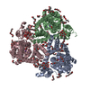
| Title | Cryo-EM structure of a substrate-bound glutamate transporter homologue GltTk encapsulated within a nanodisc | ||||||
 Components Components | Proton/glutamate symporter, SDF family | ||||||
 Keywords Keywords | TRANSPORT PROTEIN / Glutamate / transporter / mutant / nanodisc | ||||||
| Function / homology |  Function and homology information Function and homology informationdicarboxylic acid transport / symporter activity / membrane / plasma membrane Similarity search - Function | ||||||
| Biological species |   Thermococcus kodakarensis (archaea) Thermococcus kodakarensis (archaea) | ||||||
| Method | ELECTRON MICROSCOPY / single particle reconstruction / cryo EM / Resolution: 3.25 Å | ||||||
 Authors Authors | Whittaker, J.J. / Guskov, A. | ||||||
| Funding support |  Netherlands, 1items Netherlands, 1items
| ||||||
 Citation Citation |  Journal: Nat Commun / Year: 2023 Journal: Nat Commun / Year: 2023Title: Mutation in glutamate transporter homologue GltTk provides insights into pathologic mechanism of episodic ataxia 6. Authors: Emanuela Colucci / Zaid R Anshari / Miyer F Patiño-Ruiz / Mariia Nemchinova / Jacob Whittaker / Dirk J Slotboom / Albert Guskov /  Abstract: Episodic ataxias (EAs) are rare neurological conditions affecting the nervous system and typically leading to motor impairment. EA6 is linked to the mutation of a highly conserved proline into an ...Episodic ataxias (EAs) are rare neurological conditions affecting the nervous system and typically leading to motor impairment. EA6 is linked to the mutation of a highly conserved proline into an arginine in the glutamate transporter EAAT1. In vitro studies showed that this mutation leads to a reduction in the substrates transport and an increase in the anion conductance. It was hypothesised that the structural basis of these opposed functional effects might be the straightening of transmembrane helix 5, which is kinked in the wild-type protein. In this study, we present the functional and structural implications of the mutation P208R in the archaeal homologue of glutamate transporters Glt. We show that also in Glt the P208R mutation leads to reduced aspartate transport activity and increased anion conductance, however a cryo-EM structure reveals that the kink is preserved. The arginine side chain of the mutant points towards the lipidic environment, where it may engage in interactions with the phospholipids, thereby potentially interfering with the transport cycle and contributing to stabilisation of an anion conducting state. #1:  Journal: Nat Commun / Year: 2020 Journal: Nat Commun / Year: 2020Title: Structural ensemble of a glutamate transporter homologue in lipid nanodisc environment. Authors: Valentina Arkhipova / Albert Guskov / Dirk J Slotboom /   Abstract: Glutamate transporters are cation-coupled secondary active membrane transporters that clear the neurotransmitter L-glutamate from the synaptic cleft. These transporters are homotrimers, with each ...Glutamate transporters are cation-coupled secondary active membrane transporters that clear the neurotransmitter L-glutamate from the synaptic cleft. These transporters are homotrimers, with each protomer functioning independently by an elevator-type mechanism, in which a mobile transport domain alternates between inward- and outward-oriented states. Using single-particle cryo-EM we have determined five structures of the glutamate transporter homologue Glt, a Na- L-aspartate symporter, embedded in lipid nanodiscs. Dependent on the substrate concentrations used, the protomers of the trimer adopt a variety of asymmetrical conformations, consistent with the independent movement. Six of the 15 resolved protomers are in a hitherto elusive state of the transport cycle in which the inward-facing transporters are loaded with Na ions. These structures explain how substrate-leakage is prevented - a strict requirement for coupled transport. The belt protein of the lipid nanodiscs bends around the inward oriented protomers, suggesting that membrane deformations occur during transport. | ||||||
| History |
|
- Structure visualization
| Structure viewer | Molecule:  Molmil Molmil Jmol/JSmol Jmol/JSmol |
|---|
- Downloads & links
- Download
| PDBx/mmCIF format |  8afa.cif.gz 8afa.cif.gz | 227.7 KB | Display |  PDBx/mmCIF format PDBx/mmCIF format |
|---|---|---|---|---|
| PDB format |  pdb8afa.ent.gz pdb8afa.ent.gz | 181.9 KB | Display |  PDB format PDB format |
| PDBx/mmJSON format |  8afa.json.gz 8afa.json.gz | Tree view |  PDBx/mmJSON format PDBx/mmJSON format | |
| Others |  Other downloads Other downloads |
-Validation report
| Arichive directory |  https://data.pdbj.org/pub/pdb/validation_reports/af/8afa https://data.pdbj.org/pub/pdb/validation_reports/af/8afa ftp://data.pdbj.org/pub/pdb/validation_reports/af/8afa ftp://data.pdbj.org/pub/pdb/validation_reports/af/8afa | HTTPS FTP |
|---|
-Related structure data
| Related structure data |  15393MC M: map data used to model this data C: citing same article ( |
|---|---|
| Similar structure data | Similarity search - Function & homology  F&H Search F&H Search |
| Experimental dataset #1 | Data reference:  10.1038/ncomms13420 / Data set type: diffraction image data / Metadata reference: 10.1038/ncomms13420 10.1038/ncomms13420 / Data set type: diffraction image data / Metadata reference: 10.1038/ncomms13420 |
- Links
Links
- Assembly
| Deposited unit | 
| ||||||||||||||||||||||||||||||||||||||||||||||||||||||||||||||||||||||||||||||||||||||||||||||
|---|---|---|---|---|---|---|---|---|---|---|---|---|---|---|---|---|---|---|---|---|---|---|---|---|---|---|---|---|---|---|---|---|---|---|---|---|---|---|---|---|---|---|---|---|---|---|---|---|---|---|---|---|---|---|---|---|---|---|---|---|---|---|---|---|---|---|---|---|---|---|---|---|---|---|---|---|---|---|---|---|---|---|---|---|---|---|---|---|---|---|---|---|---|---|---|
| 1 |
| ||||||||||||||||||||||||||||||||||||||||||||||||||||||||||||||||||||||||||||||||||||||||||||||
| Noncrystallographic symmetry (NCS) | NCS domain:
NCS domain segments:
NCS oper:
|
- Components
| #1: Protein | Mass: 46745.441 Da / Num. of mol.: 3 / Mutation: P208R Source method: isolated from a genetically manipulated source Details: Mutant P208R / Source: (gene. exp.)   Thermococcus kodakarensis (archaea) / Strain: ATCC BAA-918 / JCM 12380 / KOD1 / Cell: E.coli / Gene: TK0986 / Production host: Thermococcus kodakarensis (archaea) / Strain: ATCC BAA-918 / JCM 12380 / KOD1 / Cell: E.coli / Gene: TK0986 / Production host:   Thermococcus kodakarensis (archaea) / References: UniProt: Q5JID0 Thermococcus kodakarensis (archaea) / References: UniProt: Q5JID0#2: Chemical | Has ligand of interest | N | |
|---|
-Experimental details
-Experiment
| Experiment | Method: ELECTRON MICROSCOPY |
|---|---|
| EM experiment | Aggregation state: PARTICLE / 3D reconstruction method: single particle reconstruction |
- Sample preparation
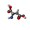
| Component | Name: Glutamate transporter homologue GltTK in a lipid nanodisc environment bound with L-aspartate ligands Type: COMPLEX / Entity ID: #1 / Source: RECOMBINANT | ||||||||||||||||||||
|---|---|---|---|---|---|---|---|---|---|---|---|---|---|---|---|---|---|---|---|---|---|
| Molecular weight | Value: 1.39 MDa / Experimental value: YES | ||||||||||||||||||||
| Source (natural) | Organism:   Thermococcus kodakarensis (archaea) / Strain: BL21(DE3) Thermococcus kodakarensis (archaea) / Strain: BL21(DE3) | ||||||||||||||||||||
| Source (recombinant) | Organism:  | ||||||||||||||||||||
| Buffer solution | pH: 8 | ||||||||||||||||||||
| Buffer component |
| ||||||||||||||||||||
| Specimen | Conc.: 1.5 mg/ml / Embedding applied: NO / Shadowing applied: NO / Staining applied: NO / Vitrification applied: YES | ||||||||||||||||||||
| Specimen support | Grid material: GOLD / Grid mesh size: 300 divisions/in. / Grid type: Quantifoil R1.2/1.3 | ||||||||||||||||||||
| Vitrification | Cryogen name: ETHANE / Humidity: 100 % |
- Electron microscopy imaging
| Experimental equipment |  Model: Titan Krios / Image courtesy: FEI Company |
|---|---|
| Microscopy | Model: FEI TITAN KRIOS |
| Electron gun | Electron source:  FIELD EMISSION GUN / Accelerating voltage: 300 kV / Illumination mode: FLOOD BEAM FIELD EMISSION GUN / Accelerating voltage: 300 kV / Illumination mode: FLOOD BEAM |
| Electron lens | Mode: BRIGHT FIELD / Nominal defocus max: 5000 nm / Nominal defocus min: 1000 nm |
| Image recording | Electron dose: 50 e/Å2 / Film or detector model: GATAN K3 (6k x 4k) |
| Image scans | Scanner model: OTHER |
- Processing
| Software | Name: PHENIX / Version: 1.20.1_4487: / Classification: refinement | ||||||||||||||||||||||||
|---|---|---|---|---|---|---|---|---|---|---|---|---|---|---|---|---|---|---|---|---|---|---|---|---|---|
| CTF correction | Type: PHASE FLIPPING AND AMPLITUDE CORRECTION | ||||||||||||||||||||||||
| Symmetry | Point symmetry: C3 (3 fold cyclic) | ||||||||||||||||||||||||
| 3D reconstruction | Resolution: 3.25 Å / Resolution method: OTHER / Num. of particles: 4576 / Symmetry type: POINT | ||||||||||||||||||||||||
| Refinement | Cross valid method: NONE Stereochemistry target values: GeoStd + Monomer Library + CDL v1.2 | ||||||||||||||||||||||||
| Displacement parameters | Biso mean: 50.94 Å2 | ||||||||||||||||||||||||
| Refine LS restraints |
| ||||||||||||||||||||||||
| Refine LS restraints NCS |
|
 Movie
Movie Controller


 PDBj
PDBj


