 Movie
Movie Controller


+ Open data

- Basic information
| Entry | Database: PDB / ID: 8acu | ||||||
|---|---|---|---|---|---|---|---|
| Title | Structure of Bacillus subtilis Rel in complex with DarB | ||||||
 Components Components |
| ||||||
 Keywords Keywords | TRANSFERASE / Rel / GTP pyrophosphokinase / DarB / CBPB / stringent factor / translation / ribosome | ||||||
| Function / homology |  Function and homology information Function and homology informationGTP diphosphokinase / GTP diphosphokinase activity / guanosine tetraphosphate biosynthetic process / kinase activity / GTP binding / ATP binding Similarity search - Function | ||||||
| Biological species |  | ||||||
| Method |  X-RAY DIFFRACTION / SYNCHROTRON / MOLECULAR REPLACEMENT / Resolution: 2.97 Å X-RAY DIFFRACTION / SYNCHROTRON / MOLECULAR REPLACEMENT / Resolution: 2.97 Å | ||||||
 Authors Authors | Garcia-Pino, A. | ||||||
| Funding support |  Belgium, 1items Belgium, 1items
| ||||||
 Citation Citation | Journal: To Be Published Title: Structure of Bacillus subtilis Rel in complex with DarB Authors: Garcia-Pino, A. | ||||||
| History |
|
- Structure visualization
| Structure viewer | Molecule: MolmilJmol/JSmol |
|---|
- Downloads & links
-Download
| PDBx/mmCIF format | 8acu.cif.gz | 380.4 KB | Display | PDBx/mmCIF format |
|---|---|---|---|---|
| PDB format | pdb8acu.ent.gz | 309.7 KB | Display | PDB format |
| PDBx/mmJSON format | 8acu.json.gz | Tree view | PDBx/mmJSON format | |
| Others |  Other downloads Other downloads |
-Validation report
| Arichive directory | https://data.pdbj.org/pub/pdb/validation_reports/ac/8acuftp://data.pdbj.org/pub/pdb/validation_reports/ac/8acu | HTTPS FTP |
|---|
-Related structure data
| Related structure data |  6s2tS S: Starting model for refinement |
|---|---|
| Similar structure data |
-Links
 PDBj
PDBj


- Assembly
| Deposited unit | 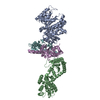
| ||||||||
|---|---|---|---|---|---|---|---|---|---|
| 1 |
| ||||||||
| Unit cell |
|
-Components
| #1: Protein | Mass: 43479.012 Da / Num. of mol.: 2 Source method: isolated from a genetically manipulated source Source: (gene. exp.) Production host: References: UniProt: O54408, GTP diphosphokinase #2: Protein | Mass: 16653.346 Da / Num. of mol.: 2 Source method: isolated from a genetically manipulated source Source: (gene. exp.) Production host: References: UniProt: A0A164SLA6 #3: Chemical |   Mass: 54.938 Da / Num. of mol.: 2 / Source method: obtained synthetically / Formula: Mn Mass: 54.938 Da / Num. of mol.: 2 / Source method: obtained synthetically / Formula: Mn#4: Water | ChemComp-HOH / |  Mass: 18.015 Da / Num. of mol.: 42 / Source method: isolated from a natural source / Formula: H2O Mass: 18.015 Da / Num. of mol.: 42 / Source method: isolated from a natural source / Formula: H2OHas ligand of interest | N | |
|---|
-Experimental details
-Experiment
| Experiment | Method: X-RAY DIFFRACTION / Number of used crystals: 1 |
|---|
- Sample preparation
| Crystal | Density Matthews: 2.65 Å3/Da / Density % sol: 53.65 % |
|---|---|
| Crystal grow | Temperature: 293.16 K / Method: vapor diffusion, sitting drop / pH: 7.5 Details: 0.1 M Magnesium chloride hexahydrate 0.1 M Sodium HEPES 7.5 10 % w/v PEG 4000 |
-Data collection
| Diffraction | Mean temperature: 100 K / Serial crystal experiment: N |
|---|---|
| Diffraction source | Source: SYNCHROTRON / Site: SOLEIL  / Beamline: PROXIMA 2 / Wavelength: 0.9801 Å / Beamline: PROXIMA 2 / Wavelength: 0.9801 Å |
| Detector | Type: DECTRIS EIGER X 16M / Detector: PIXEL / Date: Dec 20, 2020 |
| Radiation | Protocol: SINGLE WAVELENGTH / Monochromatic (M) / Laue (L): M / Scattering type: x-ray |
| Radiation wavelength | Wavelength: 0.9801 Å / Relative weight: 1 |
| Reflection | Resolution: 2.97→77.6 Å / Num. obs: 22489 / % possible obs: 95 % / Redundancy: 20.1 % / CC1/2: 0.998 / Rpim(I) all: 0.061 / Net I/σ(I): 11.1 |
| Reflection shell | Resolution: 2.97→3.08 Å / Num. unique obs: 1023 / CC1/2: 0.387 / Rpim(I) all: 0.851 |
- Processing
| Software |
| |||||||||||||||||||||||||||||||||||||||||||||||||||||||||||||||
|---|---|---|---|---|---|---|---|---|---|---|---|---|---|---|---|---|---|---|---|---|---|---|---|---|---|---|---|---|---|---|---|---|---|---|---|---|---|---|---|---|---|---|---|---|---|---|---|---|---|---|---|---|---|---|---|---|---|---|---|---|---|---|---|---|
| Refinement | Method to determine structure: MOLECULAR REPLACEMENT Starting model: 6S2T Resolution: 2.97→75.72 Å / SU ML: 0.49 / Cross valid method: THROUGHOUT / σ(F): 1.34 / Phase error: 34.52 / Stereochemistry target values: MLHL
| |||||||||||||||||||||||||||||||||||||||||||||||||||||||||||||||
| Solvent computation | Shrinkage radii: 0.9 Å / VDW probe radii: 1.1 Å / Solvent model: FLAT BULK SOLVENT MODEL | |||||||||||||||||||||||||||||||||||||||||||||||||||||||||||||||
| Refinement step | Cycle: LAST / Resolution: 2.97→75.72 Å
| |||||||||||||||||||||||||||||||||||||||||||||||||||||||||||||||
| Refine LS restraints |
| |||||||||||||||||||||||||||||||||||||||||||||||||||||||||||||||
| LS refinement shell |
| |||||||||||||||||||||||||||||||||||||||||||||||||||||||||||||||
| Refinement TLS params. | Method: refined / Origin x: 16.9521 Å / Origin y: -24.4312 Å / Origin z: 27.0744 Å
| |||||||||||||||||||||||||||||||||||||||||||||||||||||||||||||||
| Refinement TLS group | Selection details: all |
