 Movie
Movie Controller

[English] 日本語
 Yorodumi
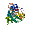
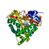
- PDB-8abo: Crystal structure of a staphylococcal orthologue of CYP134A1 (CYP... -
+ Open data

- Basic information
| Entry | Database: PDB / ID: 8abo | ||||||
|---|---|---|---|---|---|---|---|
| Title | Crystal structure of a staphylococcal orthologue of CYP134A1 (CYPX) in complex with a fragment | ||||||
 Components Components | CYPX | ||||||
 Keywords Keywords | OXIDOREDUCTASE / CYP / CYPX / Staphylococcus aureus / CLL / Pulcherremin / P450 | ||||||
| Function / homology | 4-PHENYL-1H-IMIDAZOLE / PROTOPORPHYRIN IX CONTAINING FE / NITRATE ION Function and homology information Function and homology information | ||||||
| Biological species |  Staphylococcus (bacteria) Staphylococcus (bacteria) | ||||||
| Method |  X-RAY DIFFRACTION / SYNCHROTRON / MOLECULAR REPLACEMENT / Resolution: 1.97 Å X-RAY DIFFRACTION / SYNCHROTRON / MOLECULAR REPLACEMENT / Resolution: 1.97 Å | ||||||
 Authors Authors | Snee, M. / Katariya, M. / Levy, C. | ||||||
| Funding support |  United Kingdom, 1items United Kingdom, 1items
| ||||||
 Citation Citation | Journal: To Be Published Title: Crystal structure of a staphylococcal orthologue of CYP134A1 (CYPX) in complex with a fragment Authors: Snee, M. / Katariya, M. | ||||||
| History |
|
- Structure visualization
| Structure viewer | Molecule: MolmilJmol/JSmol |
|---|
- Downloads & links
-Download
| PDBx/mmCIF format | 8abo.cif.gz | 225 KB | Display | PDBx/mmCIF format |
|---|---|---|---|---|
| PDB format | pdb8abo.ent.gz | 150.4 KB | Display | PDB format |
| PDBx/mmJSON format | 8abo.json.gz | Tree view | PDBx/mmJSON format | |
| Others |  Other downloads Other downloads |
-Validation report
| Arichive directory | https://data.pdbj.org/pub/pdb/validation_reports/ab/8aboftp://data.pdbj.org/pub/pdb/validation_reports/ab/8abo | HTTPS FTP |
|---|
-Related structure data
| Related structure data | 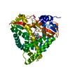 8adrC 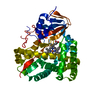 7pq1S S: Starting model for refinement C: citing same article ( |
|---|---|
| Similar structure data |
-Links
 PDBj
PDBj- Assembly
| Deposited unit | 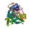
| ||||||||||||
|---|---|---|---|---|---|---|---|---|---|---|---|---|---|
| 1 |
| ||||||||||||
| Unit cell |
|
-Components
| #1: Protein | Mass: 45974.688 Da / Num. of mol.: 1 Source method: isolated from a genetically manipulated source Source: (gene. exp.) Staphylococcus (bacteria) / Plasmid: pET47b / Production host: | ||||
|---|---|---|---|---|---|
| #2: Chemical | ChemComp-231 /   Mass: 144.173 Da / Num. of mol.: 1 / Source method: obtained synthetically / Formula: C9H8N2 / Feature type: SUBJECT OF INVESTIGATION Mass: 144.173 Da / Num. of mol.: 1 / Source method: obtained synthetically / Formula: C9H8N2 / Feature type: SUBJECT OF INVESTIGATION | ||||
| #3: Chemical | ChemComp-HEM /   Mass: 616.487 Da / Num. of mol.: 1 / Source method: obtained synthetically / Formula: C34H32FeN4O4 Mass: 616.487 Da / Num. of mol.: 1 / Source method: obtained synthetically / Formula: C34H32FeN4O4 | ||||
| #4: Chemical |   Mass: 62.005 Da / Num. of mol.: 2 / Source method: obtained synthetically / Formula: NO3 Mass: 62.005 Da / Num. of mol.: 2 / Source method: obtained synthetically / Formula: NO3#5: Water | ChemComp-HOH / |  Mass: 18.015 Da / Num. of mol.: 236 / Source method: isolated from a natural source / Formula: H2O Mass: 18.015 Da / Num. of mol.: 236 / Source method: isolated from a natural source / Formula: H2OHas ligand of interest | Y | |
-Experimental details
-Experiment
| Experiment | Method: X-RAY DIFFRACTION / Number of used crystals: 1 |
|---|
- Sample preparation
| Crystal | Density Matthews: 2.32 Å3/Da / Density % sol: 47 % |
|---|---|
| Crystal grow | Temperature: 277.15 K / Method: vapor diffusion, sitting drop / pH: 8.5 Details: 0.2M Ammonium nitrate, 0.1M Bis-Tris propane pH 8.5, 18% v/v PEG smear High Temp details: 4C |
-Data collection
| Diffraction | Mean temperature: 100 K / Serial crystal experiment: N |
|---|---|
| Diffraction source | Source: SYNCHROTRON / Site: Diamond / Beamline: I03 / Wavelength: 0.9763 Å |
| Detector | Type: DECTRIS EIGER2 X 16M / Detector: PIXEL / Date: Nov 26, 2020 |
| Radiation | Protocol: SINGLE WAVELENGTH / Monochromatic (M) / Laue (L): M / Scattering type: x-ray |
| Radiation wavelength | Wavelength: 0.9763 Å / Relative weight: 1 |
| Reflection | Resolution: 1.97→53.68 Å / Num. obs: 30618 / % possible obs: 100 % / Redundancy: 12.7 % / Biso Wilson estimate: 34.01 Å2 / CC1/2: 1 / Rmerge(I) obs: 0.074 / Rpim(I) all: 0.02 / Net I/σ(I): 17.5 |
| Reflection shell | Resolution: 1.97→2.02 Å / Redundancy: 12.8 % / Rmerge(I) obs: 0.736 / Mean I/σ(I) obs: 1.3 / Num. unique obs: 2129 / CC1/2: 0.967 / Rpim(I) all: 0.213 / % possible all: 100 |
- Processing
| Software |
| ||||||||||||||||||||||||||||||||||||||||||||||||||||||||||||||||||||||||||||||||||||
|---|---|---|---|---|---|---|---|---|---|---|---|---|---|---|---|---|---|---|---|---|---|---|---|---|---|---|---|---|---|---|---|---|---|---|---|---|---|---|---|---|---|---|---|---|---|---|---|---|---|---|---|---|---|---|---|---|---|---|---|---|---|---|---|---|---|---|---|---|---|---|---|---|---|---|---|---|---|---|---|---|---|---|---|---|---|
| Refinement | Method to determine structure: MOLECULAR REPLACEMENT Starting model: 7PQ1 Resolution: 1.97→53.68 Å / SU ML: 0.2367 / Cross valid method: FREE R-VALUE / σ(F): 1.39 / Phase error: 25.0004 Stereochemistry target values: GeoStd + Monomer Library + CDL v1.2
| ||||||||||||||||||||||||||||||||||||||||||||||||||||||||||||||||||||||||||||||||||||
| Solvent computation | Shrinkage radii: 0.9 Å / VDW probe radii: 1.1 Å / Solvent model: FLAT BULK SOLVENT MODEL | ||||||||||||||||||||||||||||||||||||||||||||||||||||||||||||||||||||||||||||||||||||
| Displacement parameters | Biso mean: 47.38 Å2 | ||||||||||||||||||||||||||||||||||||||||||||||||||||||||||||||||||||||||||||||||||||
| Refinement step | Cycle: LAST / Resolution: 1.97→53.68 Å
| ||||||||||||||||||||||||||||||||||||||||||||||||||||||||||||||||||||||||||||||||||||
| Refine LS restraints |
| ||||||||||||||||||||||||||||||||||||||||||||||||||||||||||||||||||||||||||||||||||||
| LS refinement shell |
| ||||||||||||||||||||||||||||||||||||||||||||||||||||||||||||||||||||||||||||||||||||
| Refinement TLS params. | Method: refined / Origin x: -16.5499592451 Å / Origin y: -22.2090157304 Å / Origin z: -5.93775278281 Å
| ||||||||||||||||||||||||||||||||||||||||||||||||||||||||||||||||||||||||||||||||||||
| Refinement TLS group | Selection details: all |
