 Movie
Movie Controller

[English] 日本語
 Yorodumi
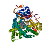
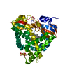
- PDB-8aa7: Crystal structure of a staphylococcal orthologue of CYP134A1 (CYP... -
+ Open data

- Basic information
| Entry | Database: PDB / ID: 8aa7 | ||||||
|---|---|---|---|---|---|---|---|
| Title | Crystal structure of a staphylococcal orthologue of CYP134A1 (CYPX) in complex with a heme coordinated fragment | ||||||
 Components Components | Cytochrome P450 protein | ||||||
 Keywords Keywords | OXIDOREDUCTASE / CYP / P450 / CYP134A1 / CLL / Cyclic / Diketopiperazine / pulcherrimin / staphylococcus / aureus | ||||||
| Function / homology |  Function and homology information Function and homology informationOxidoreductases; Acting on paired donors, with incorporation or reduction of molecular oxygen / oxidoreductase activity, acting on paired donors, with incorporation or reduction of molecular oxygen / monooxygenase activity / iron ion binding / heme binding Similarity search - Function | ||||||
| Biological species |   Staphylococcus aureus (bacteria) Staphylococcus aureus (bacteria) | ||||||
| Method |  X-RAY DIFFRACTION / SYNCHROTRON / MOLECULAR REPLACEMENT / Resolution: 2.05 Å X-RAY DIFFRACTION / SYNCHROTRON / MOLECULAR REPLACEMENT / Resolution: 2.05 Å | ||||||
 Authors Authors | Snee, M. / Katariya, M. / Levy, C. / Leys, D. | ||||||
| Funding support |  United Kingdom, 1items United Kingdom, 1items
| ||||||
 Citation Citation | Journal: To Be Published Title: Crystal structure of a staphylococcal orthologue of CYP134A1 (CYPX) in complex with a heme coordinated fragment Authors: Snee, M. / Katariya, M. | ||||||
| History |
|
- Structure visualization
| Structure viewer | Molecule: MolmilJmol/JSmol |
|---|
- Downloads & links
-Download
| PDBx/mmCIF format | 8aa7.cif.gz | 316.4 KB | Display | PDBx/mmCIF format |
|---|---|---|---|---|
| PDB format | pdb8aa7.ent.gz | 215.2 KB | Display | PDB format |
| PDBx/mmJSON format | 8aa7.json.gz | Tree view | PDBx/mmJSON format | |
| Others |  Other downloads Other downloads |
-Validation report
| Arichive directory | https://data.pdbj.org/pub/pdb/validation_reports/aa/8aa7ftp://data.pdbj.org/pub/pdb/validation_reports/aa/8aa7 | HTTPS FTP |
|---|
-Related structure data
| Related structure data | 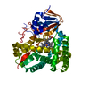 7pq1S S: Starting model for refinement |
|---|---|
| Similar structure data |
-Links
 PDBj
PDBj
- Assembly
| Deposited unit | 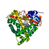
| ||||||||||||
|---|---|---|---|---|---|---|---|---|---|---|---|---|---|
| 1 |
| ||||||||||||
| Unit cell |
|
-Components
| #1: Protein | Mass: 45974.688 Da / Num. of mol.: 1 Source method: isolated from a genetically manipulated source Source: (gene. exp.) Staphylococcus aureus (bacteria) / Gene: cypX_1, NCTC5664_01270, cypX / Plasmid: pET47b / Production host: References: UniProt: A0A380DQV1, pulcherriminic acid synthase |
|---|---|
| #2: Chemical | ChemComp-WYH / 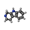  Mass: 172.226 Da / Num. of mol.: 1 / Source method: obtained synthetically / Formula: C11H12N2 / Feature type: SUBJECT OF INVESTIGATION Mass: 172.226 Da / Num. of mol.: 1 / Source method: obtained synthetically / Formula: C11H12N2 / Feature type: SUBJECT OF INVESTIGATION |
| #3: Chemical | ChemComp-HEM /   Mass: 616.487 Da / Num. of mol.: 1 / Source method: obtained synthetically / Formula: C34H32FeN4O4 Mass: 616.487 Da / Num. of mol.: 1 / Source method: obtained synthetically / Formula: C34H32FeN4O4 |
| #4: Water | ChemComp-HOH /  Mass: 18.015 Da / Num. of mol.: 255 / Source method: isolated from a natural source / Formula: H2O Mass: 18.015 Da / Num. of mol.: 255 / Source method: isolated from a natural source / Formula: H2O |
| Has ligand of interest | Y |
-Experimental details
-Experiment
| Experiment | Method: X-RAY DIFFRACTION / Number of used crystals: 1 |
|---|
- Sample preparation
| Crystal | Density Matthews: 2.45 Å3/Da / Density % sol: 49.86 % |
|---|---|
| Crystal grow | Temperature: 277.15 K / Method: vapor diffusion, sitting drop / pH: 7.8 Details: 0.05M Magnesium chloride hexahydrate, 0.05M Sodium citrate tribasic dihydrate, 0.1M Bis-Tris propane pH7.8, 22.5 % v/v PEG Smear High |
-Data collection
| Diffraction | Mean temperature: 100 K / Ambient temp details: 4C / Serial crystal experiment: N |
|---|---|
| Diffraction source | Source: SYNCHROTRON / Site: Diamond / Beamline: I03 / Wavelength: 0.9763 Å |
| Detector | Type: DECTRIS EIGER2 XE 16M / Detector: PIXEL / Date: Nov 26, 2020 |
| Radiation | Protocol: SINGLE WAVELENGTH / Monochromatic (M) / Laue (L): M / Scattering type: x-ray |
| Radiation wavelength | Wavelength: 0.9763 Å / Relative weight: 1 |
| Reflection | Resolution: 2.05→54.79 Å / Num. obs: 28754 / % possible obs: 100 % / Redundancy: 12.9 % / Biso Wilson estimate: 37.27 Å2 / CC1/2: 0.999 / Rmerge(I) obs: 0.094 / Rpim(I) all: 0.027 / Rrim(I) all: 0.098 / Net I/σ(I): 15.4 |
| Reflection shell | Resolution: 2.05→2.11 Å / Redundancy: 12.3 % / Rmerge(I) obs: 0.786 / Mean I/σ(I) obs: 1.3 / Num. unique obs: 2190 / CC1/2: 0.941 / Rpim(I) all: 0.234 / Rrim(I) all: 0.82 / % possible all: 100 |
- Processing
| Software |
| |||||||||||||||||||||||||||||||||||||||||||||||||||||||||||||||||||||||||||||
|---|---|---|---|---|---|---|---|---|---|---|---|---|---|---|---|---|---|---|---|---|---|---|---|---|---|---|---|---|---|---|---|---|---|---|---|---|---|---|---|---|---|---|---|---|---|---|---|---|---|---|---|---|---|---|---|---|---|---|---|---|---|---|---|---|---|---|---|---|---|---|---|---|---|---|---|---|---|---|
| Refinement | Method to determine structure: MOLECULAR REPLACEMENT Starting model: 7PQ1 Resolution: 2.05→47.52 Å / SU ML: 0.2294 / Cross valid method: FREE R-VALUE / σ(F): 1.39 / Phase error: 22.7751 Stereochemistry target values: GeoStd + Monomer Library + CDL v1.2
| |||||||||||||||||||||||||||||||||||||||||||||||||||||||||||||||||||||||||||||
| Solvent computation | Shrinkage radii: 0.9 Å / VDW probe radii: 1.1 Å / Solvent model: FLAT BULK SOLVENT MODEL | |||||||||||||||||||||||||||||||||||||||||||||||||||||||||||||||||||||||||||||
| Displacement parameters | Biso mean: 51.95 Å2 | |||||||||||||||||||||||||||||||||||||||||||||||||||||||||||||||||||||||||||||
| Refinement step | Cycle: LAST / Resolution: 2.05→47.52 Å
| |||||||||||||||||||||||||||||||||||||||||||||||||||||||||||||||||||||||||||||
| Refine LS restraints |
| |||||||||||||||||||||||||||||||||||||||||||||||||||||||||||||||||||||||||||||
| LS refinement shell |
| |||||||||||||||||||||||||||||||||||||||||||||||||||||||||||||||||||||||||||||
| Refinement TLS params. | Method: refined / Origin x: -17.197048431 Å / Origin y: -22.7340135464 Å / Origin z: -5.66737327573 Å
| |||||||||||||||||||||||||||||||||||||||||||||||||||||||||||||||||||||||||||||
| Refinement TLS group | Selection details: all |
