 Movie
Movie Controller

[English] 日本語
 Yorodumi
- PDB-8a83: rsEGFP2 with a chlorinated chromophore in the fluorescent ON-stat... -
+ Open data

- Basic information
| Entry | Database: PDB / ID: 8a83 | ||||||||||||
|---|---|---|---|---|---|---|---|---|---|---|---|---|---|
| Title | rsEGFP2 with a chlorinated chromophore in the fluorescent ON-state in a crystal dehydrated after illumination | ||||||||||||
 Components Components | Green fluorescent protein | ||||||||||||
 Keywords Keywords | FLUORESCENT PROTEIN / GFP-like protein / beta-barrel / bioluminescence | ||||||||||||
| Function / homology | Green fluorescent protein, GFP / Green fluorescent protein-related / Green fluorescent protein / Green fluorescent protein / bioluminescence / generation of precursor metabolites and energy / Green fluorescent protein Function and homology information Function and homology information | ||||||||||||
| Biological species |   Aequorea victoria (jellyfish) Aequorea victoria (jellyfish) | ||||||||||||
| Method |  X-RAY DIFFRACTION / SYNCHROTRON / MOLECULAR REPLACEMENT / Resolution: 1.81 Å X-RAY DIFFRACTION / SYNCHROTRON / MOLECULAR REPLACEMENT / Resolution: 1.81 Å | ||||||||||||
 Authors Authors | Fadini, A. / van Thor, J. / Chang, J. | ||||||||||||
| Funding support |  United Kingdom, United Kingdom,  United States, 3items United States, 3items
| ||||||||||||
 Citation Citation | Journal: J.Am.Chem.Soc. / Year: 2023 Title: Serial Femtosecond Crystallography Reveals that Photoactivation in a Fluorescent Protein Proceeds via the Hula Twist Mechanism. Authors: Fadini, A. / Hutchison, C.D.M. / Morozov, D. / Chang, J. / Maghlaoui, K. / Perrett, S. / Luo, F. / Kho, J.C.X. / Romei, M.G. / Morgan, R.M.L. / Orr, C.M. / Cordon-Preciado, V. / Fujiwara, T. ...Authors: Fadini, A. / Hutchison, C.D.M. / Morozov, D. / Chang, J. / Maghlaoui, K. / Perrett, S. / Luo, F. / Kho, J.C.X. / Romei, M.G. / Morgan, R.M.L. / Orr, C.M. / Cordon-Preciado, V. / Fujiwara, T. / Nuemket, N. / Tosha, T. / Tanaka, R. / Owada, S. / Tono, K. / Iwata, S. / Boxer, S.G. / Groenhof, G. / Nango, E. / van Thor, J.J. | ||||||||||||
| History |
|
- Structure visualization
| Structure viewer | Molecule: MolmilJmol/JSmol |
|---|
- Downloads & links
-Download
| PDBx/mmCIF format | 8a83.cif.gz | 63.9 KB | Display | PDBx/mmCIF format |
|---|---|---|---|---|
| PDB format | pdb8a83.ent.gz | 43.5 KB | Display | PDB format |
| PDBx/mmJSON format | 8a83.json.gz | Tree view | PDBx/mmJSON format | |
| Others |  Other downloads Other downloads |
-Validation report
| Summary document | 8a83_validation.pdf.gz | 438.6 KB | Display | wwPDB validaton report |
|---|---|---|---|---|
| Full document | 8a83_full_validation.pdf.gz | 440.6 KB | Display | |
| Data in XML | 8a83_validation.xml.gz | 12.1 KB | Display | |
| Data in CIF | 8a83_validation.cif.gz | 16.4 KB | Display | |
| Arichive directory | https://data.pdbj.org/pub/pdb/validation_reports/a8/8a83ftp://data.pdbj.org/pub/pdb/validation_reports/a8/8a83 | HTTPS FTP |
-Related structure data
| Related structure data |  8a6gC  8a6nC  8a6oC  8a6pC  8a6qC 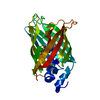 8a6rC  8a6sC 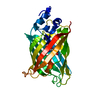 8a7vC 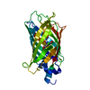 8am4C  5dtyS S: Starting model for refinement C: citing same article ( |
|---|---|
| Similar structure data |
-Links
 PDBj
PDBj

- Assembly
| Deposited unit | 
| ||||||||
|---|---|---|---|---|---|---|---|---|---|
| 1 |
| ||||||||
| Unit cell |
|
-Components
| #1: Protein | Mass: 28566.648 Da / Num. of mol.: 1 Source method: isolated from a genetically manipulated source Source: (gene. exp.) Aequorea victoria (jellyfish) / Gene: GFP / Production host:  | ||||
|---|---|---|---|---|---|
| #2: Chemical | ChemComp-SO4 /   Mass: 96.063 Da / Num. of mol.: 4 / Source method: obtained synthetically / Formula: SO4 Mass: 96.063 Da / Num. of mol.: 4 / Source method: obtained synthetically / Formula: SO4#3: Water | ChemComp-HOH / |  Mass: 18.015 Da / Num. of mol.: 109 / Source method: isolated from a natural source / Formula: H2O Mass: 18.015 Da / Num. of mol.: 109 / Source method: isolated from a natural source / Formula: H2OHas ligand of interest | Y | |
-Experimental details
-Experiment
| Experiment | Method: X-RAY DIFFRACTION / Number of used crystals: 1 |
|---|
- Sample preparation
| Crystal | Density % sol: 27.63 % |
|---|---|
| Crystal grow | Temperature: 298 K / Method: vapor diffusion, hanging drop Details: 1.76 M ammonium sulfate, 0.1 M HEPES; Cryoprotectant 1.6 M sucrose, 2.72 M ammonium sulfate, 0.16 M HEPES |
-Data collection
| Diffraction | Mean temperature: 100 K / Serial crystal experiment: N |
|---|---|
| Diffraction source | Source: SYNCHROTRON / Site: SSRL / Beamline: BL7-1 / Wavelength: 1.19 Å |
| Detector | Type: ADSC QUANTUM 315r / Detector: CCD / Date: Jun 26, 2018 |
| Radiation | Protocol: SINGLE WAVELENGTH / Monochromatic (M) / Laue (L): M / Scattering type: x-ray |
| Radiation wavelength | Wavelength: 1.19 Å / Relative weight: 1 |
| Reflection | Resolution: 1.81→38.79 Å / Num. obs: 18434 / % possible obs: 99.54 % / Redundancy: 2 % / CC1/2: 1 / Net I/σ(I): 16.16 |
| Reflection shell | Resolution: 1.81→1.875 Å / Num. unique obs: 1806 / CC1/2: 0.794 |
- Processing
| Software |
| ||||||||||||||||||||||||||||||||||||||||||||||||
|---|---|---|---|---|---|---|---|---|---|---|---|---|---|---|---|---|---|---|---|---|---|---|---|---|---|---|---|---|---|---|---|---|---|---|---|---|---|---|---|---|---|---|---|---|---|---|---|---|---|
| Refinement | Method to determine structure: MOLECULAR REPLACEMENT Starting model: 5DTY Resolution: 1.81→38.786 Å / SU ML: 0.2 / Cross valid method: THROUGHOUT / σ(F): 1.34 / Phase error: 27.99 / Stereochemistry target values: ML
| ||||||||||||||||||||||||||||||||||||||||||||||||
| Solvent computation | Shrinkage radii: 0.9 Å / VDW probe radii: 1.11 Å / Solvent model: FLAT BULK SOLVENT MODEL | ||||||||||||||||||||||||||||||||||||||||||||||||
| Displacement parameters | Biso max: 117.35 Å2 / Biso mean: 34.4947 Å2 / Biso min: 19.76 Å2 | ||||||||||||||||||||||||||||||||||||||||||||||||
| Refinement step | Cycle: final / Resolution: 1.81→38.786 Å
| ||||||||||||||||||||||||||||||||||||||||||||||||
| LS refinement shell | Refine-ID: X-RAY DIFFRACTION / Rfactor Rfree error: 0
|
