 Movie
Movie Controller

[English] 日本語
 Yorodumi
- PDB-8a7n: Crystal Structure of human Brachyury G177D variant in complex wit... -
+ Open data

- Basic information
| Entry | Database: PDB / ID: 8a7n | ||||||
|---|---|---|---|---|---|---|---|
| Title | Crystal Structure of human Brachyury G177D variant in complex with (S)-N-(3-aminopropyl)-3-((1-(2-fluorophenyl)-2-oxopyrrolidin-3-yl)amino)-N-methylbenzamide (CF-2-125) | ||||||
 Components Components | T-box transcription factor T | ||||||
 Keywords Keywords | TRANSCRIPTION / Brachyury / Chordoma / TBXT | ||||||
| Function / homology |  Function and homology information Function and homology informationprimitive streak formation / anterior/posterior axis specification, embryo / Epithelial-Mesenchymal Transition (EMT) during gastrulation / cardiac muscle cell myoblast differentiation / Germ layer formation at gastrulation / Formation of definitive endoderm / Formation of axial mesoderm / cell fate specification / Cardiogenesis / Formation of paraxial mesoderm ...primitive streak formation / anterior/posterior axis specification, embryo / Epithelial-Mesenchymal Transition (EMT) during gastrulation / cardiac muscle cell myoblast differentiation / Germ layer formation at gastrulation / Formation of definitive endoderm / Formation of axial mesoderm / cell fate specification / Cardiogenesis / Formation of paraxial mesoderm / mesoderm development / mesoderm formation / somitogenesis / heart morphogenesis / sequence-specific double-stranded DNA binding / transcription corepressor activity / RNA polymerase II-specific DNA-binding transcription factor binding / DNA-binding transcription factor activity, RNA polymerase II-specific / RNA polymerase II cis-regulatory region sequence-specific DNA binding / DNA-binding transcription factor activity / regulation of transcription by RNA polymerase II / chromatin / negative regulation of transcription by RNA polymerase II / signal transduction / positive regulation of transcription by RNA polymerase II / nucleoplasm / nucleus Similarity search - Function | ||||||
| Biological species |  Homo sapiens (human) Homo sapiens (human) | ||||||
| Method |  X-RAY DIFFRACTION / SYNCHROTRON / FOURIER SYNTHESIS / Resolution: 1.9 Å X-RAY DIFFRACTION / SYNCHROTRON / FOURIER SYNTHESIS / Resolution: 1.9 Å | ||||||
 Authors Authors | Newman, J.A. / Gavard, A. / Aitkenhead, H. / Imprachim, N. / Sherestha, L. / Burgess-Brown, N.A. / von Delft, F. / Bountra, C. / Gileadi, O. | ||||||
| Funding support |  United States, 1items United States, 1items
| ||||||
 Citation Citation | Journal: Nat Commun / Year: 2025 Title: Structural insights into human brachyury DNA recognition and discovery of progressible binders for cancer therapy. Authors: Newman, J.A. / Gavard, A.E. / Imprachim, N. / Aitkenhead, H. / Sheppard, H.E. / Te Poele, R. / Clarke, P.A. / Hossain, M.A. / Temme, L. / Oh, H.J. / Wells, C.I. / Davis-Gilbert, Z.W. / ...Authors: Newman, J.A. / Gavard, A.E. / Imprachim, N. / Aitkenhead, H. / Sheppard, H.E. / Te Poele, R. / Clarke, P.A. / Hossain, M.A. / Temme, L. / Oh, H.J. / Wells, C.I. / Davis-Gilbert, Z.W. / Workman, P. / Gileadi, O. / Drewry, D.H. #1: Journal: Acta Crystallogr.,Sect.D / Year: 2012 Title: Towards automated crystallographic structure refinement with phenix.refine. Authors: Afonine, P.V. / Grosse-Kunstleve, R.W. / Echols, N. / Headd, J.J. / Moriarty, N.W. / Mustyakimov, M. / Terwilliger, T.C. / Urzhumtsev, A. / Zwart, P.H. / Adams, P.D. #2: Journal: Acta Crystallogr D Struct Biol / Year: 2019 Title: Macromolecular structure determination using X-rays, neutrons and electrons: recent developments in Phenix. Authors: Dorothee Liebschner / Pavel V Afonine / Matthew L Baker / Gábor Bunkóczi / Vincent B Chen / Tristan I Croll / Bradley Hintze / Li Wei Hung / Swati Jain / Airlie J McCoy / Nigel W Moriarty ...Authors: Dorothee Liebschner / Pavel V Afonine / Matthew L Baker / Gábor Bunkóczi / Vincent B Chen / Tristan I Croll / Bradley Hintze / Li Wei Hung / Swati Jain / Airlie J McCoy / Nigel W Moriarty / Robert D Oeffner / Billy K Poon / Michael G Prisant / Randy J Read / Jane S Richardson / David C Richardson / Massimo D Sammito / Oleg V Sobolev / Duncan H Stockwell / Thomas C Terwilliger / Alexandre G Urzhumtsev / Lizbeth L Videau / Christopher J Williams / Paul D Adams /   Abstract: Diffraction (X-ray, neutron and electron) and electron cryo-microscopy are powerful methods to determine three-dimensional macromolecular structures, which are required to understand biological ...Diffraction (X-ray, neutron and electron) and electron cryo-microscopy are powerful methods to determine three-dimensional macromolecular structures, which are required to understand biological processes and to develop new therapeutics against diseases. The overall structure-solution workflow is similar for these techniques, but nuances exist because the properties of the reduced experimental data are different. Software tools for structure determination should therefore be tailored for each method. Phenix is a comprehensive software package for macromolecular structure determination that handles data from any of these techniques. Tasks performed with Phenix include data-quality assessment, map improvement, model building, the validation/rebuilding/refinement cycle and deposition. Each tool caters to the type of experimental data. The design of Phenix emphasizes the automation of procedures, where possible, to minimize repetitive and time-consuming manual tasks, while default parameters are chosen to encourage best practice. A graphical user interface provides access to many command-line features of Phenix and streamlines the transition between programs, project tracking and re-running of previous tasks. | ||||||
| History |
|
- Structure visualization
| Structure viewer | Molecule: MolmilJmol/JSmol |
|---|
- Downloads & links
-Download
| PDBx/mmCIF format | 8a7n.cif.gz | 66.7 KB | Display | PDBx/mmCIF format |
|---|---|---|---|---|
| PDB format | pdb8a7n.ent.gz | 38.7 KB | Display | PDB format |
| PDBx/mmJSON format | 8a7n.json.gz | Tree view | PDBx/mmJSON format | |
| Others |  Other downloads Other downloads |
-Validation report
| Arichive directory | https://data.pdbj.org/pub/pdb/validation_reports/a7/8a7nftp://data.pdbj.org/pub/pdb/validation_reports/a7/8a7n | HTTPS FTP |
|---|
-Related structure data
| Related structure data |  6f58C 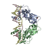 6f59C 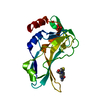 7zk2C  7zkfC  7zl2C  8a10C  8cdnC  5qt0S S: Starting model for refinement C: citing same article ( |
|---|---|
| Similar structure data |
-Links
 PDBj
PDBj


- Assembly
| Deposited unit | 
| |||||||||||||||
|---|---|---|---|---|---|---|---|---|---|---|---|---|---|---|---|---|
| 1 |
| |||||||||||||||
| Unit cell |
| |||||||||||||||
| Components on special symmetry positions |
|
-Components
| #1: Protein | Mass: 19655.623 Da / Num. of mol.: 1 Source method: isolated from a genetically manipulated source Source: (gene. exp.) Homo sapiens (human) / Gene: TBXT, T / Production host:  |
|---|---|
| #2: Chemical | ChemComp-PO4 /   Mass: 94.971 Da / Num. of mol.: 1 / Source method: obtained synthetically / Formula: PO4 Mass: 94.971 Da / Num. of mol.: 1 / Source method: obtained synthetically / Formula: PO4 |
| #3: Chemical | ChemComp-L9E / 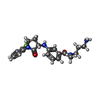  Mass: 384.447 Da / Num. of mol.: 1 / Source method: obtained synthetically / Formula: C21H25FN4O2 / Feature type: SUBJECT OF INVESTIGATION Mass: 384.447 Da / Num. of mol.: 1 / Source method: obtained synthetically / Formula: C21H25FN4O2 / Feature type: SUBJECT OF INVESTIGATION |
| #4: Water | ChemComp-HOH /  Mass: 18.015 Da / Num. of mol.: 148 / Source method: isolated from a natural source / Formula: H2O Mass: 18.015 Da / Num. of mol.: 148 / Source method: isolated from a natural source / Formula: H2O |
| Has ligand of interest | Y |
| Has protein modification | N |
-Experimental details
-Experiment
| Experiment | Method: X-RAY DIFFRACTION / Number of used crystals: 1 |
|---|
- Sample preparation
| Crystal | Density Matthews: 2.45 Å3/Da / Density % sol: 49.74 % |
|---|---|
| Crystal grow | Temperature: 277 K / Method: vapor diffusion, sitting drop / Details: 0.1 M SPG pH 7.0, 30 % PEG 1000 |
-Data collection
| Diffraction | Mean temperature: 100 K / Serial crystal experiment: N |
|---|---|
| Diffraction source | Source: SYNCHROTRON / Site: Diamond / Beamline: I03 / Wavelength: 0.97625 Å |
| Detector | Type: DECTRIS PILATUS 6M / Detector: PIXEL / Date: Jul 18, 2021 |
| Radiation | Protocol: SINGLE WAVELENGTH / Monochromatic (M) / Laue (L): M / Scattering type: x-ray |
| Radiation wavelength | Wavelength: 0.97625 Å / Relative weight: 1 |
| Reflection | Resolution: 1.9→50.2 Å / Num. obs: 15336 / % possible obs: 100 % / Redundancy: 18.4 % / Biso Wilson estimate: 32.14 Å2 / CC1/2: 1 / Rmerge(I) obs: 0.105 / Rpim(I) all: 0.025 / Net I/σ(I): 18 |
| Reflection shell | Resolution: 1.9→1.94 Å / Rmerge(I) obs: 1.782 / Mean I/σ(I) obs: 1.9 / Num. unique obs: 976 / CC1/2: 0.805 / Rpim(I) all: 0.581 / % possible all: 100 |
- Processing
| Software |
| ||||||||||||||||||||||||||||||||||||||||||
|---|---|---|---|---|---|---|---|---|---|---|---|---|---|---|---|---|---|---|---|---|---|---|---|---|---|---|---|---|---|---|---|---|---|---|---|---|---|---|---|---|---|---|---|
| Refinement | Method to determine structure: FOURIER SYNTHESIS Starting model: 5QT0 Resolution: 1.9→50.2 Å / SU ML: 0.2631 / Cross valid method: FREE R-VALUE / σ(F): 1.33 / Phase error: 29.8488 Stereochemistry target values: GeoStd + Monomer Library + CDL v1.2
| ||||||||||||||||||||||||||||||||||||||||||
| Solvent computation | Shrinkage radii: 0.9 Å / VDW probe radii: 1.11 Å / Solvent model: FLAT BULK SOLVENT MODEL | ||||||||||||||||||||||||||||||||||||||||||
| Displacement parameters | Biso mean: 38.07 Å2 | ||||||||||||||||||||||||||||||||||||||||||
| Refinement step | Cycle: LAST / Resolution: 1.9→50.2 Å
| ||||||||||||||||||||||||||||||||||||||||||
| Refine LS restraints |
| ||||||||||||||||||||||||||||||||||||||||||
| LS refinement shell |
|
