 Movie
Movie Controller

[English] 日本語
 Yorodumi
- PDB-8a6d: 10 picosecond light activated crystal structure of bovine rhodops... -
+ Open data

- Basic information
| Entry | Database: PDB / ID: 8a6d | |||||||||
|---|---|---|---|---|---|---|---|---|---|---|
| Title | 10 picosecond light activated crystal structure of bovine rhodopsin in Lipidic Cubic Phase | |||||||||
 Components Components | Rhodopsin | |||||||||
 Keywords Keywords | MEMBRANE PROTEIN / GPCR / Opsin | |||||||||
| Function / homology |  Function and homology information Function and homology informationOpsins / VxPx cargo-targeting to cilium / sperm head plasma membrane / rod bipolar cell differentiation / absorption of visible light / opsin binding / The canonical retinoid cycle in rods (twilight vision) / G protein-coupled opsin signaling pathway / 11-cis retinal binding / podosome assembly ...Opsins / VxPx cargo-targeting to cilium / sperm head plasma membrane / rod bipolar cell differentiation / absorption of visible light / opsin binding / The canonical retinoid cycle in rods (twilight vision) / G protein-coupled opsin signaling pathway / 11-cis retinal binding / podosome assembly / G protein-coupled photoreceptor activity / photoreceptor inner segment membrane / cellular response to light stimulus / rod photoreceptor outer segment / G protein-coupled receptor complex / Inactivation, recovery and regulation of the phototransduction cascade / thermotaxis / Activation of the phototransduction cascade / outer membrane / detection of temperature stimulus involved in thermoception / response to light intensity / photoreceptor cell maintenance / arrestin family protein binding / photoreceptor outer segment membrane / G alpha (i) signalling events / response to light stimulus / phototransduction, visible light / phototransduction / G-protein alpha-subunit binding / photoreceptor outer segment / sperm midpiece / visual perception / guanyl-nucleotide exchange factor activity / microtubule cytoskeleton organization / photoreceptor disc membrane / cell-cell junction / gene expression / G protein-coupled receptor signaling pathway / Golgi membrane / zinc ion binding / identical protein binding / membrane / plasma membrane Similarity search - Function | |||||||||
| Biological species |  | |||||||||
| Method |  X-RAY DIFFRACTION / FREE ELECTRON LASER / MOLECULAR REPLACEMENT / Resolution: 1.8 Å X-RAY DIFFRACTION / FREE ELECTRON LASER / MOLECULAR REPLACEMENT / Resolution: 1.8 Å | |||||||||
 Authors Authors | Gruhl, T. / Weinert, T. / Rodrigues, M.J. / Milne, C.J. / Ortolani, G. / Nass, K. / Nango, E. / Sen, S. / Johnson, P.J.M. / Cirelli, C. ...Gruhl, T. / Weinert, T. / Rodrigues, M.J. / Milne, C.J. / Ortolani, G. / Nass, K. / Nango, E. / Sen, S. / Johnson, P.J.M. / Cirelli, C. / Furrer, A. / Mous, S. / Skopintsev, P. / James, D. / Dworkowski, F. / Baath, P. / Kekilli, D. / Oserov, D. / Tanaka, R. / Glover, H. / Bacellar, C. / Bruenle, S. / Casadei, C.M. / Diethelm, A.D. / Gashi, D. / Gotthard, G. / Guixa-Gonzalez, R. / Joti, Y. / Kabanova, V. / Knopp, G. / Lesca, E. / Ma, P. / Martiel, I. / Muehle, J. / Owada, S. / Pamula, F. / Sarabi, D. / Tejero, O. / Tsai, C.J. / Varma, N. / Wach, A. / Boutet, S. / Tono, K. / Nogly, P. / Deupi, X. / Iwata, S. / Neutze, R. / Standfuss, J. / Schertler, G.F.X. / Panneels, V. | |||||||||
| Funding support |  Switzerland, 2items Switzerland, 2items
| |||||||||
 Citation Citation | Journal: Nature / Year: 2023 Title: Ultrafast structural changes direct the first molecular events of vision. Authors: Gruhl, T. / Weinert, T. / Rodrigues, M.J. / Milne, C.J. / Ortolani, G. / Nass, K. / Nango, E. / Sen, S. / Johnson, P.J.M. / Cirelli, C. / Furrer, A. / Mous, S. / Skopintsev, P. / James, D. / ...Authors: Gruhl, T. / Weinert, T. / Rodrigues, M.J. / Milne, C.J. / Ortolani, G. / Nass, K. / Nango, E. / Sen, S. / Johnson, P.J.M. / Cirelli, C. / Furrer, A. / Mous, S. / Skopintsev, P. / James, D. / Dworkowski, F. / Bath, P. / Kekilli, D. / Ozerov, D. / Tanaka, R. / Glover, H. / Bacellar, C. / Brunle, S. / Casadei, C.M. / Diethelm, A.D. / Gashi, D. / Gotthard, G. / Guixa-Gonzalez, R. / Joti, Y. / Kabanova, V. / Knopp, G. / Lesca, E. / Ma, P. / Martiel, I. / Muhle, J. / Owada, S. / Pamula, F. / Sarabi, D. / Tejero, O. / Tsai, C.J. / Varma, N. / Wach, A. / Boutet, S. / Tono, K. / Nogly, P. / Deupi, X. / Iwata, S. / Neutze, R. / Standfuss, J. / Schertler, G. / Panneels, V. #1: Journal: Biorxiv / Year: 2022Title: ULTRAFAST STRUCTURAL CHANGES DIRECT THE FIRST MOLECULAR EVENTS OF VISION Authors: Gruhl, T. / Weinert, T. / Rodrigues, M. / Milne, C.J. / Ortolani, G. / Nass, K. / Nango, E. / Sen, S. / Johnson, P.J.M. / Cirelli, C. / Furrer, A. / Mous, S. / Skopintsev, P. / James, D. / ...Authors: Gruhl, T. / Weinert, T. / Rodrigues, M. / Milne, C.J. / Ortolani, G. / Nass, K. / Nango, E. / Sen, S. / Johnson, P.J.M. / Cirelli, C. / Furrer, A. / Mous, S. / Skopintsev, P. / James, D. / Dworkowski, F. / Bath, P. / Kekilli, D. / Ozerov, D. / Tanaka, R. / Glover, H. / Bacellar, C. / Brunle, S. / Casadei, C.M. / Diethelm, A.D. / Gashi, D. / Gotthard, G. / Guixa-Gonzalez, R. / Joti, Y. / Kabanova, V. / Knopp, G. / Lesca, E. / Ma, P. / Martiel, I. / Muhle, J. / Owada, S. / Pamula, F. / Sarabi, D. / Tejero, O. / Tsai, C.J. / Varma, N. / Wach, A. / Boutet, S. / Tono, K. / Nogly, P. / Deupi, X. / Iwata, S. / Neutze, R. / Standfuss, J. / Schertler, G.F. / Panneels, V. | |||||||||
| History |
|
- Structure visualization
| Structure viewer | Molecule: MolmilJmol/JSmol |
|---|
- Downloads & links
-Download
| PDBx/mmCIF format | 8a6d.cif.gz | 335 KB | Display | PDBx/mmCIF format |
|---|---|---|---|---|
| PDB format | pdb8a6d.ent.gz | 226.5 KB | Display | PDB format |
| PDBx/mmJSON format | 8a6d.json.gz | Tree view | PDBx/mmJSON format | |
| Others |  Other downloads Other downloads |
-Validation report
| Summary document | 8a6d_validation.pdf.gz | 3.3 MB | Display | wwPDB validaton report |
|---|---|---|---|---|
| Full document | 8a6d_full_validation.pdf.gz | 3.3 MB | Display | |
| Data in XML | 8a6d_validation.xml.gz | 25.9 KB | Display | |
| Data in CIF | 8a6d_validation.cif.gz | 35.2 KB | Display | |
| Arichive directory | https://data.pdbj.org/pub/pdb/validation_reports/a6/8a6dftp://data.pdbj.org/pub/pdb/validation_reports/a6/8a6d | HTTPS FTP |
-Related structure data
| Related structure data |  7zbcC  7zbeC  8a6cC  8a6eC 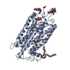 1u19S S: Starting model for refinement C: citing same article ( |
|---|---|
| Similar structure data |
-Links
 PDBj
PDBj


- Assembly
| Deposited unit | 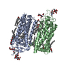
| ||||||||||||
|---|---|---|---|---|---|---|---|---|---|---|---|---|---|
| 1 | 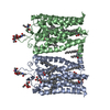
| ||||||||||||
| Unit cell |
|
-Components
-Protein , 1 types, 2 molecules AB
| #1: Protein | Mass: 39031.457 Da / Num. of mol.: 2 / Source method: isolated from a natural source / Source: (natural) |
|---|
-Sugars , 2 types, 4 molecules 
| #2: Polysaccharide | Source method: isolated from a genetically manipulated source #5: Sugar |  Type: D-saccharide, beta linking / Mass: 221.208 Da / Num. of mol.: 2 / Source method: obtained synthetically / Formula: C8H15NO6 Type: D-saccharide, beta linking / Mass: 221.208 Da / Num. of mol.: 2 / Source method: obtained synthetically / Formula: C8H15NO6 |
|---|
-Non-polymers , 6 types, 134 molecules 


| #3: Chemical |  Mass: 44.053 Da / Num. of mol.: 2 / Source method: obtained synthetically / Formula: C2H4O Mass: 44.053 Da / Num. of mol.: 2 / Source method: obtained synthetically / Formula: C2H4O#4: Chemical |  Mass: 284.436 Da / Num. of mol.: 2 / Source method: obtained synthetically / Formula: C20H28O / Feature type: SUBJECT OF INVESTIGATION Mass: 284.436 Da / Num. of mol.: 2 / Source method: obtained synthetically / Formula: C20H28O / Feature type: SUBJECT OF INVESTIGATION#6: Chemical | ChemComp-DAO / |  Mass: 200.318 Da / Num. of mol.: 1 / Source method: obtained synthetically / Formula: C12H24O2 Mass: 200.318 Da / Num. of mol.: 1 / Source method: obtained synthetically / Formula: C12H24O2#7: Chemical | ChemComp-OLC / (  Mass: 356.540 Da / Num. of mol.: 13 / Source method: obtained synthetically / Formula: C21H40O4 Mass: 356.540 Da / Num. of mol.: 13 / Source method: obtained synthetically / Formula: C21H40O4#8: Chemical |  Mass: 256.424 Da / Num. of mol.: 2 / Source method: obtained synthetically / Formula: C16H32O2 Mass: 256.424 Da / Num. of mol.: 2 / Source method: obtained synthetically / Formula: C16H32O2#9: Water | ChemComp-HOH / | Mass: 18.015 Da / Num. of mol.: 114 / Source method: isolated from a natural source / Formula: H2O |
|---|
-Details
| Has ligand of interest | Y |
|---|
-Experimental details
-Experiment
| Experiment | Method: X-RAY DIFFRACTION / Number of used crystals: 1 |
|---|
- Sample preparation
| Crystal | Density Matthews: 2.68 Å3/Da / Density % sol: 54.15 % |
|---|---|
| Crystal grow | Temperature: 294 K / Method: lipidic cubic phase / pH: 9 / Details: 36 % PEG 600, 100 mM Bicine pH 9.0 |
-Data collection
| Diffraction | Mean temperature: 294 K / Serial crystal experiment: Y |
|---|---|
| Diffraction source | Source: FREE ELECTRON LASER / Site: SwissFEL ARAMIS / Beamline: ESA / Wavelength: 1.38 Å |
| Detector | Type: PSI JUNGFRAU 1M / Detector: PIXEL / Date: Jul 24, 2020 |
| Radiation | Protocol: SINGLE WAVELENGTH / Monochromatic (M) / Laue (L): M / Scattering type: x-ray |
| Radiation wavelength | Wavelength: 1.38 Å / Relative weight: 1 |
| Reflection | Resolution: 1.8→16.1 Å / Num. obs: 79031 / % possible obs: 99.7 % / Redundancy: 595.9 % / Biso Wilson estimate: 30.44 Å2 / CC1/2: 0.9915 / CC star: 0.9979 / Net I/σ(I): 5.7 |
| Reflection shell | Resolution: 1.8→1.86 Å / Mean I/σ(I) obs: 0.72 / Num. unique obs: 79031 / CC1/2: 0.5081 / CC star: 0.8209 / R split: 1.3878 |
| Serial crystallography sample delivery | Method: injection |
- Processing
| Software |
| ||||||||||||||||||||||||||||||||||||||||||||||||||||||||
|---|---|---|---|---|---|---|---|---|---|---|---|---|---|---|---|---|---|---|---|---|---|---|---|---|---|---|---|---|---|---|---|---|---|---|---|---|---|---|---|---|---|---|---|---|---|---|---|---|---|---|---|---|---|---|---|---|---|
| Refinement | Method to determine structure: MOLECULAR REPLACEMENT Starting model: 1U19 Resolution: 1.8→9.99 Å / SU ML: 0.4175 / Cross valid method: FREE R-VALUE / Phase error: 51.1655 Stereochemistry target values: GeoStd + Monomer Library + CDL v1.2
| ||||||||||||||||||||||||||||||||||||||||||||||||||||||||
| Solvent computation | Shrinkage radii: 0.9 Å / VDW probe radii: 1.1 Å / Solvent model: FLAT BULK SOLVENT MODEL | ||||||||||||||||||||||||||||||||||||||||||||||||||||||||
| Displacement parameters | Biso mean: 44.69 Å2 | ||||||||||||||||||||||||||||||||||||||||||||||||||||||||
| Refinement step | Cycle: LAST / Resolution: 1.8→9.99 Å
| ||||||||||||||||||||||||||||||||||||||||||||||||||||||||
| Refine LS restraints |
| ||||||||||||||||||||||||||||||||||||||||||||||||||||||||
| LS refinement shell |
| ||||||||||||||||||||||||||||||||||||||||||||||||||||||||
| Refinement TLS params. | Method: refined / Origin x: -5.42370360115 Å / Origin y: 28.3745861112 Å / Origin z: 37.7684672506 Å
| ||||||||||||||||||||||||||||||||||||||||||||||||||||||||
| Refinement TLS group | Selection details: all |
