 Movie
Movie Controller

[English] 日本語
 Yorodumi
- PDB-8a67: Branched Lys48- and Lys63-linked tri-ubiquitin (K48-K63-Ub3) in c... -
+ Open data

- Basic information
| Entry | Database: PDB / ID: 8a67 | ||||||||||||
|---|---|---|---|---|---|---|---|---|---|---|---|---|---|
| Title | Branched Lys48- and Lys63-linked tri-ubiquitin (K48-K63-Ub3) in complex with matured synthetic nanobody NbSL3.3Q (3rd generation) | ||||||||||||
 Components Components |
| ||||||||||||
 Keywords Keywords | SIGNALING PROTEIN / Branched Ubiquitin / Nanobody / complex | ||||||||||||
| Function / homology |  Function and homology information Function and homology information | ||||||||||||
| Biological species |  Homo sapiens (human) Homo sapiens (human)synthetic construct (others) | ||||||||||||
| Method |  X-RAY DIFFRACTION / SYNCHROTRON / MOLECULAR REPLACEMENT / Resolution: 1.86 Å X-RAY DIFFRACTION / SYNCHROTRON / MOLECULAR REPLACEMENT / Resolution: 1.86 Å | ||||||||||||
 Authors Authors | Lange, S.M. / Kulathu, Y. | ||||||||||||
| Funding support |  United Kingdom, European Union, 3items United Kingdom, European Union, 3items
| ||||||||||||
 Citation Citation | Journal: Nat.Struct.Mol.Biol. / Year: 2024 Title: VCP/p97-associated proteins are binders and debranching enzymes of K48-K63-branched ubiquitin chains. Authors: Lange, S.M. / McFarland, M.R. / Lamoliatte, F. / Carroll, T. / Krshnan, L. / Perez-Rafols, A. / Kwasna, D. / Shen, L. / Wallace, I. / Cole, I. / Armstrong, L.A. / Knebel, A. / Johnson, C. / ...Authors: Lange, S.M. / McFarland, M.R. / Lamoliatte, F. / Carroll, T. / Krshnan, L. / Perez-Rafols, A. / Kwasna, D. / Shen, L. / Wallace, I. / Cole, I. / Armstrong, L.A. / Knebel, A. / Johnson, C. / De Cesare, V. / Kulathu, Y. #1: Journal: Biorxiv / Year: 2023Title: Comprehensive approach to study branched ubiquitin chains reveals roles for K48-K63 branches in VCP/p97-related processes Authors: Lange, S.M. / McFarland, M.R. / Lamoliatte, F. / Kwasna, D. / Shen, L. / Wallace, I. / Cole, I. / Armstrong, L.A. / Knebel, A. / Johnson, C. / De Cesare, V. / Kulathu, Y. #2: Journal: Acta Crystallogr D Biol Crystallogr / Year: 2010 Title: PHENIX: a comprehensive Python-based system for macromolecular structure solution. Authors: Paul D Adams / Pavel V Afonine / Gábor Bunkóczi / Vincent B Chen / Ian W Davis / Nathaniel Echols / Jeffrey J Headd / Li-Wei Hung / Gary J Kapral / Ralf W Grosse-Kunstleve / Airlie J McCoy ...Authors: Paul D Adams / Pavel V Afonine / Gábor Bunkóczi / Vincent B Chen / Ian W Davis / Nathaniel Echols / Jeffrey J Headd / Li-Wei Hung / Gary J Kapral / Ralf W Grosse-Kunstleve / Airlie J McCoy / Nigel W Moriarty / Robert Oeffner / Randy J Read / David C Richardson / Jane S Richardson / Thomas C Terwilliger / Peter H Zwart /  Abstract: Macromolecular X-ray crystallography is routinely applied to understand biological processes at a molecular level. However, significant time and effort are still required to solve and complete many ...Macromolecular X-ray crystallography is routinely applied to understand biological processes at a molecular level. However, significant time and effort are still required to solve and complete many of these structures because of the need for manual interpretation of complex numerical data using many software packages and the repeated use of interactive three-dimensional graphics. PHENIX has been developed to provide a comprehensive system for macromolecular crystallographic structure solution with an emphasis on the automation of all procedures. This has relied on the development of algorithms that minimize or eliminate subjective input, the development of algorithms that automate procedures that are traditionally performed by hand and, finally, the development of a framework that allows a tight integration between the algorithms. #3: Journal: Acta Crystallogr D Biol Crystallogr / Year: 2011 Title: Data processing and analysis with the autoPROC toolbox. Authors: Vonrhein, C. / Flensburg, C. / Keller, P. / Sharff, A. / Smart, O. / Paciorek, W. / Womack, T. / Bricogne, G. | ||||||||||||
| History |
|
- Structure visualization
| Structure viewer | Molecule: MolmilJmol/JSmol |
|---|
- Downloads & links
-Download
| PDBx/mmCIF format | 8a67.cif.gz | 196.8 KB | Display | PDBx/mmCIF format |
|---|---|---|---|---|
| PDB format | pdb8a67.ent.gz | 124.2 KB | Display | PDB format |
| PDBx/mmJSON format | 8a67.json.gz | Tree view | PDBx/mmJSON format | |
| Others |  Other downloads Other downloads |
-Validation report
| Arichive directory | https://data.pdbj.org/pub/pdb/validation_reports/a6/8a67ftp://data.pdbj.org/pub/pdb/validation_reports/a6/8a67 | HTTPS FTP |
|---|
-Related structure data
| Related structure data |  7nbbSC  7npoC S: Starting model for refinement C: citing same article ( |
|---|---|
| Similar structure data |
-Links
 PDBj
PDBj


- Assembly
| Deposited unit | 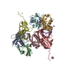
| ||||||||||||
|---|---|---|---|---|---|---|---|---|---|---|---|---|---|
| 1 | 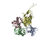
| ||||||||||||
| 2 | 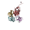
| ||||||||||||
| Unit cell |
|
-Components
-Polyubiquitin- ... , 2 types, 6 molecules AEBCFG
| #1: Protein | Mass: 8192.375 Da / Num. of mol.: 2 Source method: isolated from a genetically manipulated source Details: Ubiquitin with C-terminal truncation / Source: (gene. exp.) Homo sapiens (human) / Gene: UBB / Production host:  #2: Protein | Mass: 8632.859 Da / Num. of mol.: 4 / Mutation: K48R K63R Source method: isolated from a genetically manipulated source Source: (gene. exp.) Homo sapiens (human) / Gene: UBB / Production host: |
|---|
-Antibody , 1 types, 2 molecules DH
| #3: Antibody | Mass: 15967.914 Da / Num. of mol.: 2 Source method: isolated from a genetically manipulated source Details: Matured Nanobody NbSL3.3Q with N-terminal pelB signal sequence for periplasmic expression and C-terminal 6His affinity tag Source: (gene. exp.) synthetic construct (others) / Production host: |
|---|
-Non-polymers , 6 types, 532 molecules 


| #4: Chemical |  Mass: 92.094 Da / Num. of mol.: 2 / Source method: obtained synthetically / Formula: C3H8O3 Mass: 92.094 Da / Num. of mol.: 2 / Source method: obtained synthetically / Formula: C3H8O3#5: Chemical |  Mass: 60.095 Da / Num. of mol.: 2 / Source method: obtained synthetically / Formula: C3H8O Mass: 60.095 Da / Num. of mol.: 2 / Source method: obtained synthetically / Formula: C3H8O#6: Chemical |  Mass: 65.409 Da / Num. of mol.: 2 / Source method: obtained synthetically / Formula: Zn Mass: 65.409 Da / Num. of mol.: 2 / Source method: obtained synthetically / Formula: Zn#7: Chemical | ChemComp-CL / |  Mass: 35.453 Da / Num. of mol.: 1 / Source method: obtained synthetically / Formula: Cl Mass: 35.453 Da / Num. of mol.: 1 / Source method: obtained synthetically / Formula: Cl#8: Chemical |  Mass: 22.990 Da / Num. of mol.: 2 / Source method: obtained synthetically / Formula: Na Mass: 22.990 Da / Num. of mol.: 2 / Source method: obtained synthetically / Formula: Na#9: Water | ChemComp-HOH / | Mass: 18.015 Da / Num. of mol.: 523 / Source method: isolated from a natural source / Formula: H2O |
|---|
-Details
| Has ligand of interest | N |
|---|---|
| Has protein modification | Y |
-Experimental details
-Experiment
| Experiment | Method: X-RAY DIFFRACTION / Number of used crystals: 1 |
|---|
- Sample preparation
| Crystal | Density Matthews: 2.24 Å3/Da / Density % sol: 45.01 % |
|---|---|
| Crystal grow | Temperature: 293.15 K / Method: vapor diffusion, sitting drop / pH: 7.5 Details: Protein concentrated to 14.5 mg/ml in 20 mM HEPES pH 7.5, 150 mM NaCl. Mixed 200 nl protein with 100 nl mother liquor (0.1 M HEPES pH 7.5, 10% 2-propanol, 20% PEG4000). Crystals harvested ...Details: Protein concentrated to 14.5 mg/ml in 20 mM HEPES pH 7.5, 150 mM NaCl. Mixed 200 nl protein with 100 nl mother liquor (0.1 M HEPES pH 7.5, 10% 2-propanol, 20% PEG4000). Crystals harvested and cryo-protected with Mother liquor supplemented with 30% glycerol. |
-Data collection
| Diffraction | Mean temperature: 100 K / Serial crystal experiment: N |
|---|---|
| Diffraction source | Source: SYNCHROTRON / Site: ESRF  / Beamline: ID23-2 / Wavelength: 0.87313 Å / Beamline: ID23-2 / Wavelength: 0.87313 Å |
| Detector | Type: DECTRIS PILATUS3 2M / Detector: PIXEL / Date: Jun 9, 2022 |
| Radiation | Protocol: SINGLE WAVELENGTH / Monochromatic (M) / Laue (L): M / Scattering type: x-ray |
| Radiation wavelength | Wavelength: 0.87313 Å / Relative weight: 1 |
| Reflection | Resolution: 1.86→56.58 Å / Num. obs: 36500 / % possible obs: 88.7 % / Redundancy: 3.2 % / Biso Wilson estimate: 13.75 Å2 / CC1/2: 0.987 / Net I/σ(I): 4.1 |
| Reflection shell | Resolution: 1.86→2.09 Å / Num. unique obs: 1826 / CC1/2: 0.735 |
- Processing
| Software |
| |||||||||||||||||||||||||||||||||||||||||||||||||||||||||||||||||||||||||||||||||||||||||||
|---|---|---|---|---|---|---|---|---|---|---|---|---|---|---|---|---|---|---|---|---|---|---|---|---|---|---|---|---|---|---|---|---|---|---|---|---|---|---|---|---|---|---|---|---|---|---|---|---|---|---|---|---|---|---|---|---|---|---|---|---|---|---|---|---|---|---|---|---|---|---|---|---|---|---|---|---|---|---|---|---|---|---|---|---|---|---|---|---|---|---|---|---|
| Refinement | Method to determine structure: MOLECULAR REPLACEMENT Starting model: 7NBB Resolution: 1.86→52.59 Å / SU ML: 0.2054 / Cross valid method: FREE R-VALUE / σ(F): 1.96 / Phase error: 28.1262 Stereochemistry target values: GeoStd + Monomer Library + CDL v1.2
| |||||||||||||||||||||||||||||||||||||||||||||||||||||||||||||||||||||||||||||||||||||||||||
| Solvent computation | Shrinkage radii: 0.9 Å / VDW probe radii: 1.1 Å / Solvent model: FLAT BULK SOLVENT MODEL | |||||||||||||||||||||||||||||||||||||||||||||||||||||||||||||||||||||||||||||||||||||||||||
| Displacement parameters | Biso mean: 17.36 Å2 | |||||||||||||||||||||||||||||||||||||||||||||||||||||||||||||||||||||||||||||||||||||||||||
| Refinement step | Cycle: LAST / Resolution: 1.86→52.59 Å
| |||||||||||||||||||||||||||||||||||||||||||||||||||||||||||||||||||||||||||||||||||||||||||
| Refine LS restraints |
| |||||||||||||||||||||||||||||||||||||||||||||||||||||||||||||||||||||||||||||||||||||||||||
| LS refinement shell |
|
