[English] 日本語
Yorodumi
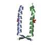
- PDB-8a50: Crystal structure of HSF2BP-ALPHA1 tetramer -

+
Open data


ID or keywords:

Loading...

-
Basic information

Entry
Database: PDB / ID: 8a50
TitleCrystal structure of HSF2BP-ALPHA1 tetramer
ComponentsHeat shock factor 2-binding protein
KeywordsRECOMBINATION / Complex
Function / homology
Function and homology information


double-strand break repair involved in meiotic recombination / female meiosis I / male meiosis I / chromosome / spermatogenesis / transcription by RNA polymerase II / nucleoplasm / cytosol
Similarity search - Function
Heat shock factor 2-binding protein / Armadillo-like helical / Armadillo-type fold
Similarity search - Domain/homology
PHOSPHATE ION / Heat shock factor 2-binding protein
Similarity search - Component
Biological speciessynthetic construct (others)
MethodX-RAY DIFFRACTION / SYNCHROTRON / MOLECULAR REPLACEMENT / Resolution: 1.484 Å
AuthorsMiron, S. / Legrand, P. / Ropars, V. / Ghouil, R. / Zinn-Justin, S.
Funding support France, 1items
OrganizationGrant numberCountry
Agence Nationale de la Recherche (ANR) France
CitationJournal: Sci Adv / Year: 2023
Title: BRCA2-HSF2BP oligomeric ring disassembly by BRME1 promotes homologous recombination.
Authors: Rania Ghouil / Simona Miron / Koichi Sato / Dejan Ristic / Sari E van Rossum-Fikkert / Pierre Legrand / Malika Ouldali / Jean-Marie Winter / Virginie Ropars / Gabriel David / Ana-Andreea ...Authors: Rania Ghouil / Simona Miron / Koichi Sato / Dejan Ristic / Sari E van Rossum-Fikkert / Pierre Legrand / Malika Ouldali / Jean-Marie Winter / Virginie Ropars / Gabriel David / Ana-Andreea Arteni / Claire Wyman / Puck Knipscheer / Roland Kanaar / Alex N Zelensky / Sophie Zinn-Justin /
Abstract: In meiotic homologous recombination (HR), BRCA2 facilitates loading of the recombinases RAD51 and DMC1 at the sites of double-strand breaks (DSBs). The HSF2BP-BRME1 complex interacts with BRCA2. Its ...In meiotic homologous recombination (HR), BRCA2 facilitates loading of the recombinases RAD51 and DMC1 at the sites of double-strand breaks (DSBs). The HSF2BP-BRME1 complex interacts with BRCA2. Its absence causes a severe reduction in recombinase loading at meiotic DSB. We previously showed that, in somatic cancer cells ectopically producing HSF2BP, DNA damage can trigger HSF2BP-dependent degradation of BRCA2, which prevents HR. Here, we report that, upon binding to BRCA2, HSF2BP forms octameric rings that are able to interlock into a large ring-shaped 24-mer. Addition of BRME1 leads to dissociation of both of these ring structures and cancels the disruptive effect of HSF2BP on cancer cell resistance to DNA damage. It also prevents BRCA2 degradation during interstrand DNA crosslink repair in egg extracts. We propose that, during meiosis, the control of HSF2BPBRCA2 oligomerization by BRME1 ensures timely assembly of the ring complex that concentrates BRCA2 and controls its turnover, thus promoting HR.
History
DepositionJun 13, 2022Deposition site: PDBE / Processing site: PDBE
Revision 1.0Jul 5, 2023Provider: repository / Type: Initial release
Revision 1.1Nov 8, 2023Group: Data collection / Database references
Category: chem_comp_atom / chem_comp_bond ...chem_comp_atom / chem_comp_bond / citation / citation_author
Item: _citation.country / _citation.journal_abbrev ..._citation.country / _citation.journal_abbrev / _citation.journal_id_CSD / _citation.journal_id_ISSN / _citation.journal_volume / _citation.page_first / _citation.page_last / _citation.pdbx_database_id_DOI / _citation.pdbx_database_id_PubMed / _citation.title / _citation.year
Revision 1.2May 1, 2024Group: Refinement description / Category: pdbx_initial_refinement_model

-
Structure visualization

Structure viewerMolecule:
MolmilJmol/JSmol

Downloads & links

-
Assembly

Deposited unit
A: Heat shock factor 2-binding protein
B: Heat shock factor 2-binding protein
hetero molecules


Theoretical massNumber of molelcules
Total (without water)7,9514
Polymers7,7612
Non-polymers1902
Water1,00956
1
A: Heat shock factor 2-binding protein
B: Heat shock factor 2-binding protein
hetero molecules

A: Heat shock factor 2-binding protein
B: Heat shock factor 2-binding protein
hetero molecules


Theoretical massNumber of molelcules
Total (without water)15,9028
Polymers15,5224
Non-polymers3804
Water724
TypeNameSymmetry operationNumber
identity operation1_555x,y,z1
crystal symmetry operation19_444-x-1/4,-z-1/4,-y-1/41
Buried area6470 Å2
ΔGint-62 kcal/mol
Surface area7220 Å2
MethodPISA
Unit cell
Length a, b, c (Å)79.81, 79.81, 79.81
Angle α, β, γ (deg.)90, 90, 90
Int Tables number213
Space group name H-MP4132
Components on special symmetry positions
IDModelComponents
11A-101-

PO4

21A-101-

PO4

-
Components

#1: Protein/peptide Heat shock factor 2-binding protein


Mass: 3880.580 Da / Num. of mol.: 2 / Source method: obtained synthetically / Source: (synth.) synthetic construct (others) / References: UniProt: O75031
#2: Chemical ChemComp-PO4 / PHOSPHATE ION


Mass: 94.971 Da / Num. of mol.: 2 / Source method: obtained synthetically / Formula: PO4
#3: Water ChemComp-HOH / water


Mass: 18.015 Da / Num. of mol.: 56 / Source method: isolated from a natural source / Formula: H2O
Has ligand of interestN

-
Experimental details

-
Experiment

ExperimentMethod: X-RAY DIFFRACTION / Number of used crystals: 1

-
Sample preparation

CrystalDensity Matthews: 2.73 Å3/Da / Density % sol: 54.93 %
Crystal growTemperature: 277 K / Method: vapor diffusion, sitting drop
Details: 0.2 M LiCl 0.1 M sodium acetate trihydrate pH 5 20% PEG6000

-
Data collection

DiffractionMean temperature: 100 K / Serial crystal experiment: N
Diffraction sourceSource: SYNCHROTRON / Site: ESRF / Beamline: MASSIF-1 / Wavelength: 0.965459 Å
DetectorType: DECTRIS PILATUS3 2M / Detector: PIXEL / Date: Nov 10, 2021 / Details: Be CRL
RadiationMonochromator: Diamond [110] / Protocol: SINGLE WAVELENGTH / Monochromatic (M) / Laue (L): M / Scattering type: x-ray
Radiation wavelengthWavelength: 0.965459 Å / Relative weight: 1
ReflectionResolution: 1.48→56.43 Å / Num. obs: 15006 / % possible obs: 99.8 % / Redundancy: 6.6 % / Biso Wilson estimate: 29.4 Å2 / CC1/2: 0.999 / Rmerge(I) obs: 0.05 / Rpim(I) all: 0.021 / Rrim(I) all: 0.054 / Net I/σ(I): 15
Reflection shellResolution: 1.48→1.52 Å / Redundancy: 6.8 % / Rmerge(I) obs: 1.393 / Mean I/σ(I) obs: 1.2 / Num. unique obs: 1084 / CC1/2: 0.559 / Rpim(I) all: 0.565 / Rrim(I) all: 1.506 / % possible all: 99.9

-
Processing

Software
NameVersionClassification
BUSTER2.10.4 (3-FEB-2022)refinement
XDSJan 10, 2022 BUILT=20220110data reduction
autoPROCdata processing
MOLREPphasing
STARANISO2.3.77data scaling
RefinementMethod to determine structure: MOLECULAR REPLACEMENT
Starting model: AlphaFold2 model

Resolution: 1.484→56.43 Å / Cor.coef. Fo:Fc: 0.946 / Cor.coef. Fo:Fc free: 0.95 / SU R Cruickshank DPI: 0.087 / Cross valid method: THROUGHOUT / SU R Blow DPI: 0.083 / SU Rfree Blow DPI: 0.075 / SU Rfree Cruickshank DPI: 0.071
RfactorNum. reflection% reflectionSelection details
Rfree0.2166 664 -RANDOM
Rwork0.2127 ---
obs0.2129 14159 94.7 %-
Displacement parametersBiso mean: 35.8 Å2
Baniso -1Baniso -2Baniso -3
1-0 Å20 Å20 Å2
2--0 Å20 Å2
3---0 Å2
Refine analyzeLuzzati coordinate error obs: 0.22 Å
Refinement stepCycle: LAST / Resolution: 1.484→56.43 Å
ProteinNucleic acidLigandSolventTotal
Num. atoms506 0 10 56 572
Refine LS restraints
Refine-IDTypeDev idealNumberRestraint functionWeight
X-RAY DIFFRACTIONt_bond_d0.0081276HARMONIC2
X-RAY DIFFRACTIONt_angle_deg0.942332HARMONIC2
X-RAY DIFFRACTIONt_dihedral_angle_d407SINUSOIDAL2
X-RAY DIFFRACTIONt_gen_planes191HARMONIC5
X-RAY DIFFRACTIONt_it1276HARMONIC10
X-RAY DIFFRACTIONt_chiral_improper_torsion78SEMIHARMONIC5
X-RAY DIFFRACTIONt_sum_occupancies9HARMONIC1
X-RAY DIFFRACTIONt_ideal_dist_contact1219SEMIHARMONIC4
X-RAY DIFFRACTIONt_omega_torsion2.94
X-RAY DIFFRACTIONt_other_torsion13.42
LS refinement shellResolution: 1.484→1.52 Å
RfactorNum. reflection% reflection
Rfree0.2707 19 -
Rwork0.2343 --
obs--46.36 %
Refinement TLS params.Origin x: -10.747 Å / Origin y: 3.7246 Å / Origin z: -15.8911 Å
111213212223313233
T-0.0073 Å20.0087 Å20.0286 Å2--0.0067 Å20.0104 Å2---0.0299 Å2
L0.3014 °2-0.213 °2-0.5051 °2-1.9523 °2-0.515 °2--0.5864 °2
S0.0803 Å °0.0348 Å °0.001 Å °0.0348 Å °0.0007 Å °0.0612 Å °0.001 Å °0.0612 Å °-0.081 Å °
Refinement TLS group
IDRefine-IDRefine TLS-IDSelection detailsAuth asym-IDAuth seq-ID
1X-RAY DIFFRACTION1{ *|* }A1 - 29
2X-RAY DIFFRACTION1{ *|* }B1 - 30
3X-RAY DIFFRACTION1{ *|* }C3 - 4
4X-RAY DIFFRACTION1{ *|* }S1 - 56

+
About Yorodumi

-
News

-
Feb 9, 2022. New format data for meta-information of EMDB entries

New format data for meta-information of EMDB entries

  • Version 3 of the EMDB header file is now the official format.
  • The previous official version 1.9 will be removed from the archive.

Related info.:EMDB header

External links:wwPDB to switch to version 3 of the EMDB data model

-
Aug 12, 2020. Covid-19 info

Covid-19 info

URL: https://pdbjlvh1.pdbj.org/emnavi/covid19.php

New page: Covid-19 featured information page in EM Navigator.

Related info.:Covid-19 info / Mar 5, 2020. Novel coronavirus structure data

+
Mar 5, 2020. Novel coronavirus structure data

Novel coronavirus structure data

Related info.:Yorodumi Speices / Aug 12, 2020. Covid-19 info

External links:COVID-19 featured content - PDBj / Molecule of the Month (242):Coronavirus Proteases

+
Jan 31, 2019. EMDB accession codes are about to change! (news from PDBe EMDB page)

EMDB accession codes are about to change! (news from PDBe EMDB page)

  • The allocation of 4 digits for EMDB accession codes will soon come to an end. Whilst these codes will remain in use, new EMDB accession codes will include an additional digit and will expand incrementally as the available range of codes is exhausted. The current 4-digit format prefixed with “EMD-” (i.e. EMD-XXXX) will advance to a 5-digit format (i.e. EMD-XXXXX), and so on. It is currently estimated that the 4-digit codes will be depleted around Spring 2019, at which point the 5-digit format will come into force.
  • The EM Navigator/Yorodumi systems omit the EMD- prefix.

Related info.:Q: What is EMD? / ID/Accession-code notation in Yorodumi/EM Navigator

External links:EMDB Accession Codes are Changing Soon! / Contact to PDBj

+
Jul 12, 2017. Major update of PDB

Major update of PDB

  • wwPDB released updated PDB data conforming to the new PDBx/mmCIF dictionary.
  • This is a major update changing the version number from 4 to 5, and with Remediation, in which all the entries are updated.
  • In this update, many items about electron microscopy experimental information are reorganized (e.g. em_software).
  • Now, EM Navigator and Yorodumi are based on the updated data.

External links:wwPDB Remediation / Enriched Model Files Conforming to OneDep Data Standards Now Available in the PDB FTP Archive

-
Yorodumi

Thousand views of thousand structures

  • Yorodumi is a browser for structure data from EMDB, PDB, SASBDB, etc.
  • This page is also the successor to EM Navigator detail page, and also detail information page/front-end page for Omokage search.
  • The word "yorodu" (or yorozu) is an old Japanese word meaning "ten thousand". "mi" (miru) is to see.

Related info.:EMDB / PDB / SASBDB / Comparison of 3 databanks / Yorodumi Search / Aug 31, 2016. New EM Navigator & Yorodumi / Yorodumi Papers / Jmol/JSmol / Function and homology information / Changes in new EM Navigator and Yorodumi

Read more