 Movie
Movie Controller


+ Open data

- Basic information
| Entry | Database: PDB / ID: 8a47 | ||||||
|---|---|---|---|---|---|---|---|
| Title | IdeS in complex with IgG1 Fc | ||||||
 Components Components |
| ||||||
 Keywords Keywords | IMMUNE SYSTEM / Antibody / protease / IgG / Fc / IdeS | ||||||
| Function / homology | Ig protease IdeS / Mac 1 / Papain-like cysteine peptidase superfamily / peptidase activity / proteolysis / IgG-degrading protease Function and homology information Function and homology information | ||||||
| Biological species |  Homo sapiens (human) Homo sapiens (human) Streptococcus pyogenes serotype M59 (bacteria) Streptococcus pyogenes serotype M59 (bacteria) | ||||||
| Method |  X-RAY DIFFRACTION / SYNCHROTRON / MOLECULAR REPLACEMENT / Resolution: 2.338 Å X-RAY DIFFRACTION / SYNCHROTRON / MOLECULAR REPLACEMENT / Resolution: 2.338 Å | ||||||
 Authors Authors | Sudol, A.S.L. / Tews, I. / Crispin, M. | ||||||
| Funding support |  United Kingdom, 1items United Kingdom, 1items
| ||||||
 Citation Citation | Journal: Nat Commun / Year: 2022 Title: Extensive substrate recognition by the streptococcal antibody-degrading enzymes IdeS and EndoS. Authors: Sudol, A.S.L. / Butler, J. / Ivory, D.P. / Tews, I. / Crispin, M. | ||||||
| History |
|
- Structure visualization
| Structure viewer | Molecule: MolmilJmol/JSmol |
|---|
- Downloads & links
-Download
| PDBx/mmCIF format | 8a47.cif.gz | 308 KB | Display | PDBx/mmCIF format |
|---|---|---|---|---|
| PDB format | pdb8a47.ent.gz | Display | PDB format | |
| PDBx/mmJSON format | 8a47.json.gz | Tree view | PDBx/mmJSON format | |
| Others |  Other downloads Other downloads |
-Validation report
| Summary document | 8a47_validation.pdf.gz | 1.4 MB | Display | wwPDB validaton report |
|---|---|---|---|---|
| Full document | 8a47_full_validation.pdf.gz | 1.4 MB | Display | |
| Data in XML | 8a47_validation.xml.gz | 28.2 KB | Display | |
| Data in CIF | 8a47_validation.cif.gz | 40.6 KB | Display | |
| Arichive directory | https://data.pdbj.org/pub/pdb/validation_reports/a4/8a47ftp://data.pdbj.org/pub/pdb/validation_reports/a4/8a47 | HTTPS FTP |
-Related structure data
| Related structure data | 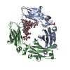 8a48C  8a49C 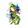 1y08S S: Starting model for refinement C: citing same article ( |
|---|---|
| Similar structure data |
-Links
 PDBj
PDBj

- Assembly
| Deposited unit | 
| ||||||||||||
|---|---|---|---|---|---|---|---|---|---|---|---|---|---|
| 1 |
| ||||||||||||
| Unit cell |
| ||||||||||||
| Noncrystallographic symmetry (NCS) | NCS domain:
NCS domain segments: Component-ID: 1 / Ens-ID: 1 / Beg auth comp-ID: PRO / Beg label comp-ID: PRO / End auth comp-ID: LEU / End label comp-ID: LEU / Refine code: 1 / Auth asym-ID: A / Label asym-ID: A / Auth seq-ID: 230 - 443 / Label seq-ID: 10 - 223
NCS ensembles : (Details: Local NCS retraints between domains: 1 2) |
-Components
-Antibody / Protein , 2 types, 3 molecules ABC
| #1: Antibody | Mass: 25554.982 Da / Num. of mol.: 2 / Mutation: E382A Source method: isolated from a genetically manipulated source Source: (gene. exp.) Homo sapiens (human) / Plasmid: pFUSE-hIgG1-Fc / Cell line (production host): HEK 293-F / Production host: Homo sapiens (human)#2: Protein | | Mass: 34930.137 Da / Num. of mol.: 1 / Mutation: C94A Source method: isolated from a genetically manipulated source Source: (gene. exp.) Streptococcus pyogenes serotype M59 (bacteria)Gene: mac, SAMEA1711581_00766 / Plasmid: pET-21a(+) / Production host: |
|---|
-Sugars , 2 types, 2 molecules
| #3: Polysaccharide | 2-acetamido-2-deoxy-beta-D-glucopyranose-(1-2)-alpha-D-mannopyranose-(1-6)-[alpha-D-mannopyranose- ...2-acetamido-2-deoxy-beta-D-glucopyranose-(1-2)-alpha-D-mannopyranose-(1-6)-[alpha-D-mannopyranose-(1-3)]beta-D-mannopyranose-(1-4)-2-acetamido-2-deoxy-beta-D-glucopyranose-(1-4)-[alpha-L-fucopyranose-(1-6)]2-acetamido-2-deoxy-beta-D-glucopyranose Type: oligosaccharide / Mass: 1260.157 Da / Num. of mol.: 1 Source method: isolated from a genetically manipulated source |
|---|---|
| #4: Polysaccharide | 2-acetamido-2-deoxy-beta-D-glucopyranose-(1-2)-alpha-D-mannopyranose-(1-3)-[2-acetamido-2-deoxy- ...2-acetamido-2-deoxy-beta-D-glucopyranose-(1-2)-alpha-D-mannopyranose-(1-3)-[2-acetamido-2-deoxy-beta-D-glucopyranose-(1-2)-alpha-D-mannopyranose-(1-6)]beta-D-mannopyranose-(1-4)-2-acetamido-2-deoxy-beta-D-glucopyranose-(1-4)-[alpha-L-fucopyranose-(1-6)]2-acetamido-2-deoxy-beta-D-glucopyranose Type: oligosaccharide / Mass: 1463.349 Da / Num. of mol.: 1 Source method: isolated from a genetically manipulated source |
-Non-polymers , 2 types, 201 molecules 

| #5: Chemical | ChemComp-NA /  Mass: 22.990 Da / Num. of mol.: 1 / Source method: obtained synthetically / Formula: Na Mass: 22.990 Da / Num. of mol.: 1 / Source method: obtained synthetically / Formula: Na |
|---|---|
| #6: Water | ChemComp-HOH / Mass: 18.015 Da / Num. of mol.: 200 / Source method: isolated from a natural source / Formula: H2O |
-Details
| Has ligand of interest | Y |
|---|---|
| Has protein modification | Y |
-Experimental details
-Experiment
| Experiment | Method: X-RAY DIFFRACTION / Number of used crystals: 1 |
|---|
- Sample preparation
| Crystal | Density Matthews: 4.08 Å3/Da / Density % sol: 69.86 % |
|---|---|
| Crystal grow | Temperature: 294.15 K / Method: vapor diffusion, sitting drop / pH: 8.5 Details: 0.12 M monosaccharides mix (0.2M D-Glucose; 0.2M D-Mannose; 0.2M D-Galactose; 0.2M L-Fucose; 0.2M D- Xylose; 0.2M N-Acetyl-D-Glucosamine), 0.1 M buffer system 3 (Tris (base); BICINE; pH 8.5) ...Details: 0.12 M monosaccharides mix (0.2M D-Glucose; 0.2M D-Mannose; 0.2M D-Galactose; 0.2M L-Fucose; 0.2M D- Xylose; 0.2M N-Acetyl-D-Glucosamine), 0.1 M buffer system 3 (Tris (base); BICINE; pH 8.5), 30 % v/v precipitant mix 1 (40% v/v PEG 500* MME; 20 % w/v PEG 20000) |
-Data collection
| Diffraction | Mean temperature: 100 K / Serial crystal experiment: N | ||||||||||||||||||
|---|---|---|---|---|---|---|---|---|---|---|---|---|---|---|---|---|---|---|---|
| Diffraction source | Source: SYNCHROTRON / Site: ESRF  / Beamline: MASSIF-3 / Wavelength: 0.9677 Å / Beamline: MASSIF-3 / Wavelength: 0.9677 Å | ||||||||||||||||||
| Detector | Type: DECTRIS EIGER X 4M / Detector: PIXEL / Date: Apr 23, 2021 / Details: Vertical CRL | ||||||||||||||||||
| Radiation | Monochromator: Si(111) / Protocol: SINGLE WAVELENGTH / Monochromatic (M) / Laue (L): M / Scattering type: x-ray | ||||||||||||||||||
| Radiation wavelength | Wavelength: 0.9677 Å / Relative weight: 1 | ||||||||||||||||||
| Reflection twin |
| ||||||||||||||||||
| Reflection | Resolution: 2.338→48.53 Å / Num. obs: 60351 / % possible obs: 97.7 % / Redundancy: 4.96 % / Rmerge(I) obs: 0.229 / Rpim(I) all: 0.112 / Rrim(I) all: 0.255 / Χ2: 0.954 / Net I/σ(I): 5.3 | ||||||||||||||||||
| Reflection shell | Resolution: 2.34→2.38 Å / Redundancy: 5 % / Rmerge(I) obs: 0.877 / Mean I/σ(I) obs: 1 / Num. unique obs: 3005 / CC1/2: 0.335 / Rpim(I) all: 0.426 / Rrim(I) all: 0.978 / % possible all: 98.8 |
- Processing
| Software |
| |||||||||||||||||||||||||||||||||||||||||||||||||||||||||||||||||||||||||||||||||||||||||||||||||||||||||||||||||||||||||||||||||||||||||||||||||||||||||||||||||||||
|---|---|---|---|---|---|---|---|---|---|---|---|---|---|---|---|---|---|---|---|---|---|---|---|---|---|---|---|---|---|---|---|---|---|---|---|---|---|---|---|---|---|---|---|---|---|---|---|---|---|---|---|---|---|---|---|---|---|---|---|---|---|---|---|---|---|---|---|---|---|---|---|---|---|---|---|---|---|---|---|---|---|---|---|---|---|---|---|---|---|---|---|---|---|---|---|---|---|---|---|---|---|---|---|---|---|---|---|---|---|---|---|---|---|---|---|---|---|---|---|---|---|---|---|---|---|---|---|---|---|---|---|---|---|---|---|---|---|---|---|---|---|---|---|---|---|---|---|---|---|---|---|---|---|---|---|---|---|---|---|---|---|---|---|---|---|---|
| Refinement | Method to determine structure: MOLECULAR REPLACEMENT Starting model: 1Y08 Resolution: 2.338→48.529 Å / Cor.coef. Fo:Fc: 0.95 / Cor.coef. Fo:Fc free: 0.927 / WRfactor Rfree: 0.18 / WRfactor Rwork: 0.153 / SU B: 2.221 / SU ML: 0.057 / Average fsc free: 0.986 / Average fsc work: 0.9914 / Cross valid method: FREE R-VALUE / ESU R: 0.038 / ESU R Free: 0.034 Details: Hydrogens have been added in their riding positions
| |||||||||||||||||||||||||||||||||||||||||||||||||||||||||||||||||||||||||||||||||||||||||||||||||||||||||||||||||||||||||||||||||||||||||||||||||||||||||||||||||||||
| Solvent computation | Ion probe radii: 0.8 Å / Shrinkage radii: 0.8 Å / VDW probe radii: 1.2 Å / Solvent model: MASK BULK SOLVENT | |||||||||||||||||||||||||||||||||||||||||||||||||||||||||||||||||||||||||||||||||||||||||||||||||||||||||||||||||||||||||||||||||||||||||||||||||||||||||||||||||||||
| Displacement parameters | Biso mean: 36.18 Å2
| |||||||||||||||||||||||||||||||||||||||||||||||||||||||||||||||||||||||||||||||||||||||||||||||||||||||||||||||||||||||||||||||||||||||||||||||||||||||||||||||||||||
| Refinement step | Cycle: LAST / Resolution: 2.338→48.529 Å
| |||||||||||||||||||||||||||||||||||||||||||||||||||||||||||||||||||||||||||||||||||||||||||||||||||||||||||||||||||||||||||||||||||||||||||||||||||||||||||||||||||||
| Refine LS restraints |
| |||||||||||||||||||||||||||||||||||||||||||||||||||||||||||||||||||||||||||||||||||||||||||||||||||||||||||||||||||||||||||||||||||||||||||||||||||||||||||||||||||||
| Refine LS restraints NCS |
| |||||||||||||||||||||||||||||||||||||||||||||||||||||||||||||||||||||||||||||||||||||||||||||||||||||||||||||||||||||||||||||||||||||||||||||||||||||||||||||||||||||
| LS refinement shell |
|
