 Movie
Movie Controller

[English] 日本語
 Yorodumi
- PDB-8a3b: Cryo-EM structure of mouse Pannexin 1 purified in Salipro nanopar... -
+ Open data

- Basic information
| Entry | Database: PDB / ID: 8a3b | ||||||
|---|---|---|---|---|---|---|---|
| Title | Cryo-EM structure of mouse Pannexin 1 purified in Salipro nanoparticles | ||||||
 Components Components | Pannexin-1 | ||||||
 Keywords Keywords | MEMBRANE PROTEIN / Membrane transporter / ATP-release channel / inflammation / immune function | ||||||
| Function / homology |  Function and homology information Function and homology informationElectric Transmission Across Gap Junctions / The NLRP3 inflammasome / ATP transmembrane transporter activity / ATP transport / leak channel activity / positive regulation of interleukin-1 alpha production / monoatomic anion transmembrane transport / wide pore channel activity / bleb / monoatomic anion channel activity ...Electric Transmission Across Gap Junctions / The NLRP3 inflammasome / ATP transmembrane transporter activity / ATP transport / leak channel activity / positive regulation of interleukin-1 alpha production / monoatomic anion transmembrane transport / wide pore channel activity / bleb / monoatomic anion channel activity / gap junction / gap junction channel activity / response to ATP / monoatomic cation transport / response to ischemia / positive regulation of cytokine production / positive regulation of interleukin-1 beta production / calcium channel activity / calcium ion transport / actin filament binding / cell-cell signaling / actin binding / protease binding / scaffold protein binding / transmembrane transporter binding / signaling receptor binding / endoplasmic reticulum membrane / protein-containing complex binding / structural molecule activity / endoplasmic reticulum / protein-containing complex / identical protein binding / plasma membrane Similarity search - Function | ||||||
| Biological species |  | ||||||
| Method | ELECTRON MICROSCOPY / single particle reconstruction / cryo EM / Resolution: 3.1 Å | ||||||
 Authors Authors | Drulyte, I. | ||||||
| Funding support | 1items
| ||||||
 Citation Citation | Journal: Sci Rep / Year: 2023 Title: Direct cell extraction of membrane proteins for structure-function analysis. Authors: Ieva Drulyte / Aspen Rene Gutgsell / Pilar Lloris-Garcerá / Michael Liss / Stefan Geschwindner / Mazdak Radjainia / Jens Frauenfeld / Robin Löving /    Abstract: Membrane proteins are the largest group of therapeutic targets in a variety of disease areas and yet, they remain particularly difficult to investigate. We have developed a novel one-step approach ...Membrane proteins are the largest group of therapeutic targets in a variety of disease areas and yet, they remain particularly difficult to investigate. We have developed a novel one-step approach for the incorporation of membrane proteins directly from cells into lipid Salipro nanoparticles. Here, with the pannexin1 channel as a case study, we demonstrate the applicability of this method for structure-function analysis using SPR and cryo-EM. | ||||||
| History |
|
- Structure visualization
| Structure viewer | Molecule: MolmilJmol/JSmol |
|---|
- Downloads & links
-Download
| PDBx/mmCIF format | 8a3b.cif.gz | 329.2 KB | Display | PDBx/mmCIF format |
|---|---|---|---|---|
| PDB format | pdb8a3b.ent.gz | 264.7 KB | Display | PDB format |
| PDBx/mmJSON format | 8a3b.json.gz | Tree view | PDBx/mmJSON format | |
| Others |  Other downloads Other downloads |
-Validation report
| Arichive directory | https://data.pdbj.org/pub/pdb/validation_reports/a3/8a3bftp://data.pdbj.org/pub/pdb/validation_reports/a3/8a3b | HTTPS FTP |
|---|
-Related structure data
| Related structure data |  15110MC M: map data used to model this data C: citing same article ( |
|---|---|
| Similar structure data |
-Links
 PDBj
PDBj- Assembly
| Deposited unit | 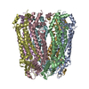
|
|---|---|
| 1 |
|
-Components
| #1: Protein | Mass: 50475.434 Da / Num. of mol.: 7 Source method: isolated from a genetically manipulated source Source: (gene. exp.)  Homo sapiens (human) / Strain (production host): Expi293 / References: UniProt: Q9JIP4 Homo sapiens (human) / Strain (production host): Expi293 / References: UniProt: Q9JIP4 |
|---|
-Experimental details
-Experiment
| Experiment | Method: ELECTRON MICROSCOPY |
|---|---|
| EM experiment | Aggregation state: PARTICLE / 3D reconstruction method: single particle reconstruction |
- Sample preparation
| Component | Name: Salipro-mPANX1 / Type: ORGANELLE OR CELLULAR COMPONENT / Entity ID: all / Source: RECOMBINANT |
|---|---|
| Molecular weight | Value: 0.353 MDa / Experimental value: NO |
| Source (natural) | Organism: |
| Source (recombinant) | Organism: Homo sapiens (human) / Strain: Expi293 |
| Buffer solution | pH: 7.5 / Details: 50 mM HEPES at pH 7.5, 150 mM NaCl |
| Specimen | Conc.: 2.5 mg/ml / Embedding applied: NO / Shadowing applied: NO / Staining applied: NO / Vitrification applied: YES |
| Specimen support | Details: Grids were glow-discharged using 20 mAmp current for 45 sec and charge set to positive. Grid material: COPPER / Grid mesh size: 200 divisions/in. / Grid type: Quantifoil R1.2/1.3 |
| Vitrification | Instrument: FEI VITROBOT MARK IV / Cryogen name: ETHANE / Humidity: 95 % / Chamber temperature: 277.15 K Details: To overcome preferred orientation, 0.5 mM fluorinated Fos-Choline 8 was added to the sample just before the grid freezing. Blot parameter: blot force +20, blot time 10 s, waiting time 30s |
- Electron microscopy imaging
| Experimental equipment |  Model: Titan Krios / Image courtesy: FEI Company |
|---|---|
| Microscopy | Model: FEI TITAN KRIOS Details: Titan Krios G4 was used with fringe-free imaging and aberration-free image shifts. Nominal pixel size for 165kx 0.75 A, calibrated pixel size 0.727 A. |
| Electron gun | Electron source:  FIELD EMISSION GUN / Accelerating voltage: 300 kV / Illumination mode: FLOOD BEAM FIELD EMISSION GUN / Accelerating voltage: 300 kV / Illumination mode: FLOOD BEAM |
| Electron lens | Mode: BRIGHT FIELD / Nominal magnification: 165000 X / Nominal defocus max: 2000 nm / Nominal defocus min: 500 nm / Cs: 2.7 mm / C2 aperture diameter: 70 µm |
| Specimen holder | Cryogen: NITROGEN / Specimen holder model: FEI TITAN KRIOS AUTOGRID HOLDER |
| Image recording | Average exposure time: 4.16 sec. / Electron dose: 40.24 e/Å2 / Film or detector model: FEI FALCON IV (4k x 4k) / Num. of grids imaged: 1 / Num. of real images: 7955 |
| EM imaging optics | Energyfilter name: TFS Selectris X / Energyfilter slit width: 10 eV |
- Processing
| EM software |
| ||||||||||||||||||||||||||||||||||||||||||||
|---|---|---|---|---|---|---|---|---|---|---|---|---|---|---|---|---|---|---|---|---|---|---|---|---|---|---|---|---|---|---|---|---|---|---|---|---|---|---|---|---|---|---|---|---|---|
| CTF correction | Type: PHASE FLIPPING AND AMPLITUDE CORRECTION | ||||||||||||||||||||||||||||||||||||||||||||
| Particle selection | Num. of particles selected: 1314251 | ||||||||||||||||||||||||||||||||||||||||||||
| Symmetry | Point symmetry: C7 (7 fold cyclic) | ||||||||||||||||||||||||||||||||||||||||||||
| 3D reconstruction | Resolution: 3.1 Å / Resolution method: FSC 0.143 CUT-OFF / Num. of particles: 268823 / Num. of class averages: 1 / Symmetry type: POINT | ||||||||||||||||||||||||||||||||||||||||||||
| Atomic model building | Protocol: RIGID BODY FIT / Space: RECIPROCAL |
