 Movie
Movie Controller

[English] 日本語
 Yorodumi
- PDB-8a0z: Crystal structure of Candida auris dihydrofolate reductase comple... -
+ Open data

- Basic information
| Entry | Database: PDB / ID: 8a0z | ||||||
|---|---|---|---|---|---|---|---|
| Title | Crystal structure of Candida auris dihydrofolate reductase complexed with NADPH and pyrimethamine | ||||||
 Components Components | Dihydrofolate reductase | ||||||
 Keywords Keywords | HYDROLASE / Dihydrofolate reductase candida auris NADPH pyrimethamine | ||||||
| Function / homology |  Function and homology information Function and homology informationdihydrofolate metabolic process / dihydrofolate reductase / dihydrofolate reductase activity / folic acid metabolic process / tetrahydrofolate biosynthetic process / one-carbon metabolic process / NADP binding / mitochondrion Similarity search - Function | ||||||
| Biological species |  [Candida] auris (fungus) [Candida] auris (fungus) | ||||||
| Method |  X-RAY DIFFRACTION / SYNCHROTRON / MOLECULAR REPLACEMENT / Resolution: 1.7 Å X-RAY DIFFRACTION / SYNCHROTRON / MOLECULAR REPLACEMENT / Resolution: 1.7 Å | ||||||
 Authors Authors | Kirkman, T.K. / Dias, M.V.B. | ||||||
| Funding support |  United Kingdom, 1items United Kingdom, 1items
| ||||||
 Citation Citation | Journal: To Be Published Title: Crystal structure of candida auris Authors: Kirkman, T.K. / Dias, M.V.B. | ||||||
| History |
|
- Structure visualization
| Structure viewer | Molecule: MolmilJmol/JSmol |
|---|
- Downloads & links
-Download
| PDBx/mmCIF format | 8a0z.cif.gz | 109.6 KB | Display | PDBx/mmCIF format |
|---|---|---|---|---|
| PDB format | pdb8a0z.ent.gz | 82.5 KB | Display | PDB format |
| PDBx/mmJSON format | 8a0z.json.gz | Tree view | PDBx/mmJSON format | |
| Others |  Other downloads Other downloads |
-Validation report
| Arichive directory | https://data.pdbj.org/pub/pdb/validation_reports/a0/8a0zftp://data.pdbj.org/pub/pdb/validation_reports/a0/8a0z | HTTPS FTP |
|---|
-Related structure data
| Related structure data |  7zzxS S: Starting model for refinement |
|---|---|
| Similar structure data |
-Links
 PDBj
PDBj

- Assembly
| Deposited unit | 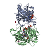
| ||||||||
|---|---|---|---|---|---|---|---|---|---|
| 1 | 
| ||||||||
| 2 | 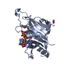
| ||||||||
| Unit cell |
|
-Components
-Protein , 1 types, 2 molecules BA
| #1: Protein | Mass: 23634.137 Da / Num. of mol.: 2 Source method: isolated from a genetically manipulated source Source: (gene. exp.) [Candida] auris (fungus) / Gene: QG37_02791 / Production host:  |
|---|
-Non-polymers , 5 types, 364 molecules 
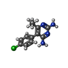


| #2: Chemical |  Mass: 745.421 Da / Num. of mol.: 2 / Source method: obtained synthetically / Formula: C21H30N7O17P3 / Feature type: SUBJECT OF INVESTIGATION Mass: 745.421 Da / Num. of mol.: 2 / Source method: obtained synthetically / Formula: C21H30N7O17P3 / Feature type: SUBJECT OF INVESTIGATION#3: Chemical |  Mass: 248.711 Da / Num. of mol.: 2 / Source method: obtained synthetically / Formula: C12H13ClN4 / Feature type: SUBJECT OF INVESTIGATION Mass: 248.711 Da / Num. of mol.: 2 / Source method: obtained synthetically / Formula: C12H13ClN4 / Feature type: SUBJECT OF INVESTIGATION#4: Chemical |  Mass: 62.005 Da / Num. of mol.: 2 / Source method: obtained synthetically / Formula: NO3 Mass: 62.005 Da / Num. of mol.: 2 / Source method: obtained synthetically / Formula: NO3#5: Chemical |  Mass: 238.305 Da / Num. of mol.: 2 / Source method: obtained synthetically / Formula: C8H18N2O4S / Comment: pH buffer*YM Mass: 238.305 Da / Num. of mol.: 2 / Source method: obtained synthetically / Formula: C8H18N2O4S / Comment: pH buffer*YM#6: Water | ChemComp-HOH / | Mass: 18.015 Da / Num. of mol.: 356 / Source method: isolated from a natural source / Formula: H2O |
|---|
-Details
| Has ligand of interest | Y |
|---|
-Experimental details
-Experiment
| Experiment | Method: X-RAY DIFFRACTION / Number of used crystals: 1 |
|---|
- Sample preparation
| Crystal | Density Matthews: 2.06 Å3/Da / Density % sol: 40.34 % |
|---|---|
| Crystal grow | Temperature: 293 K / Method: vapor diffusion, hanging drop / Details: 0.2 M Sodium nitrate, 20% PEG 3350 |
-Data collection
| Diffraction | Mean temperature: 100 K / Serial crystal experiment: N | ||||||||||||||||||||||||||||||
|---|---|---|---|---|---|---|---|---|---|---|---|---|---|---|---|---|---|---|---|---|---|---|---|---|---|---|---|---|---|---|---|
| Diffraction source | Source: SYNCHROTRON / Site: Diamond / Beamline: I04 / Wavelength: 1 Å | ||||||||||||||||||||||||||||||
| Detector | Type: DECTRIS EIGER X 16M / Detector: PIXEL / Date: Dec 9, 2021 | ||||||||||||||||||||||||||||||
| Radiation | Protocol: SINGLE WAVELENGTH / Monochromatic (M) / Laue (L): M / Scattering type: x-ray | ||||||||||||||||||||||||||||||
| Radiation wavelength | Wavelength: 1 Å / Relative weight: 1 | ||||||||||||||||||||||||||||||
| Reflection | Resolution: 1.7→51.98 Å / Num. obs: 34623 / % possible obs: 83.2 % / Redundancy: 3.5 % / CC1/2: 0.998 / Rmerge(I) obs: 0.05 / Rpim(I) all: 0.031 / Rrim(I) all: 0.059 / Net I/σ(I): 11.9 / Num. measured all: 121303 / Scaling rejects: 172 | ||||||||||||||||||||||||||||||
| Reflection shell | Diffraction-ID: 1
|
- Processing
| Software |
| |||||||||||||||||||||||||||||||||||||||||||||||||||||||||||||||||||||||||||||||||||||||||||||||||||||||||
|---|---|---|---|---|---|---|---|---|---|---|---|---|---|---|---|---|---|---|---|---|---|---|---|---|---|---|---|---|---|---|---|---|---|---|---|---|---|---|---|---|---|---|---|---|---|---|---|---|---|---|---|---|---|---|---|---|---|---|---|---|---|---|---|---|---|---|---|---|---|---|---|---|---|---|---|---|---|---|---|---|---|---|---|---|---|---|---|---|---|---|---|---|---|---|---|---|---|---|---|---|---|---|---|---|---|---|
| Refinement | Method to determine structure: MOLECULAR REPLACEMENT Starting model: 7ZZX Resolution: 1.7→41.1 Å / SU ML: 0.17 / Cross valid method: THROUGHOUT / σ(F): 1.98 / Phase error: 27 / Stereochemistry target values: ML
| |||||||||||||||||||||||||||||||||||||||||||||||||||||||||||||||||||||||||||||||||||||||||||||||||||||||||
| Solvent computation | Shrinkage radii: 0.9 Å / VDW probe radii: 1.11 Å / Solvent model: FLAT BULK SOLVENT MODEL | |||||||||||||||||||||||||||||||||||||||||||||||||||||||||||||||||||||||||||||||||||||||||||||||||||||||||
| Displacement parameters | Biso max: 59.33 Å2 / Biso mean: 22.1853 Å2 / Biso min: 8.24 Å2 | |||||||||||||||||||||||||||||||||||||||||||||||||||||||||||||||||||||||||||||||||||||||||||||||||||||||||
| Refinement step | Cycle: final / Resolution: 1.7→41.1 Å
| |||||||||||||||||||||||||||||||||||||||||||||||||||||||||||||||||||||||||||||||||||||||||||||||||||||||||
| LS refinement shell | Refine-ID: X-RAY DIFFRACTION / Rfactor Rfree error: 0 / Total num. of bins used: 14
|
