 Movie
Movie Controller

[English] 日本語
 Yorodumi
- PDB-7zwl: Crystal structure of human STING in complex with 3',3'-c-di-(2'F,... -
+ Open data

- Basic information
| Entry | Database: PDB / ID: 7zwl | ||||||||||||||||||||||||||||||
|---|---|---|---|---|---|---|---|---|---|---|---|---|---|---|---|---|---|---|---|---|---|---|---|---|---|---|---|---|---|---|---|
| Title | Crystal structure of human STING in complex with 3',3'-c-di-(2'F,2'd Components Components
 Keywords KeywordsANTIVIRAL PROTEIN / sting / antiviral / activator | Function / homology |  Function and homology information Function and homology informationSUMO is conjugated to E1 (UBA2:SAE1) / SUMOylation of nuclear envelope proteins / SUMO is transferred from E1 to E2 (UBE2I, UBC9) / SUMO is proteolytically processed / SUMOylation of transcription factors / Postmitotic nuclear pore complex (NPC) reformation / SUMOylation of transcription cofactors / septin ring / SUMOylation of DNA damage response and repair proteins / Transcriptional and post-translational regulation of MITF-M expression and activity ...SUMO is conjugated to E1 (UBA2:SAE1) / SUMOylation of nuclear envelope proteins / SUMO is transferred from E1 to E2 (UBE2I, UBC9) / SUMO is proteolytically processed / SUMOylation of transcription factors / Postmitotic nuclear pore complex (NPC) reformation / SUMOylation of transcription cofactors / septin ring / SUMOylation of DNA damage response and repair proteins / Transcriptional and post-translational regulation of MITF-M expression and activity / SUMOylation of DNA replication proteins / 2',3'-cyclic GMP-AMP binding / SUMOylation of SUMOylation proteins / Recruitment and ATM-mediated phosphorylation of repair and signaling proteins at DNA double strand breaks / SUMOylation of RNA binding proteins / SUMOylation of chromatin organization proteins / ubiquitin-like protein ligase binding / autophagosome membrane / protein sumoylation / positive regulation of type I interferon production / endoplasmic reticulum-Golgi intermediate compartment membrane / activation of innate immune response / condensed nuclear chromosome / protein tag activity / mitochondrial outer membrane / Golgi membrane / endoplasmic reticulum membrane / perinuclear region of cytoplasm / identical protein binding / nucleus Similarity search - Function Biological species |  Homo sapiens (human) Homo sapiens (human) Method |  X-RAY DIFFRACTION / SYNCHROTRON / MOLECULAR REPLACEMENT / Resolution: 2 Å X-RAY DIFFRACTION / SYNCHROTRON / MOLECULAR REPLACEMENT / Resolution: 2 Å  Authors AuthorsKlima, M. / Smola, M. / Boura, E. | Funding support | 1items |
 CitationJournal: To Be Published CitationJournal: To Be PublishedTitle: Crystal structure of human STING in complex with 3',3'-c-di-(2'F,2'd Authors: Klima, M. / Smola, M. / Boura, E. History |
|
- Structure visualization
| Structure viewer | Molecule: MolmilJmol/JSmol |
|---|
- Downloads & links
-Download
| PDBx/mmCIF format | 7zwl.cif.gz | 116.1 KB | Display | PDBx/mmCIF format |
|---|---|---|---|---|
| PDB format | pdb7zwl.ent.gz | 80.9 KB | Display | PDB format |
| PDBx/mmJSON format | 7zwl.json.gz | Tree view | PDBx/mmJSON format | |
| Others |  Other downloads Other downloads |
-Validation report
| Summary document | 7zwl_validation.pdf.gz | 908.8 KB | Display | wwPDB validaton report |
|---|---|---|---|---|
| Full document | 7zwl_full_validation.pdf.gz | 913.9 KB | Display | |
| Data in XML | 7zwl_validation.xml.gz | 18.5 KB | Display | |
| Data in CIF | 7zwl_validation.cif.gz | 26.1 KB | Display | |
| Arichive directory | https://data.pdbj.org/pub/pdb/validation_reports/zw/7zwlftp://data.pdbj.org/pub/pdb/validation_reports/zw/7zwl | HTTPS FTP |
-Related structure data
| Related structure data |  4ksyS S: Starting model for refinement |
|---|---|
| Similar structure data |
-Links
 PDBj
PDBj


- Assembly
| Deposited unit | 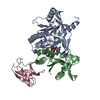
| ||||||||||||
|---|---|---|---|---|---|---|---|---|---|---|---|---|---|
| 1 |
| ||||||||||||
| Unit cell |
| ||||||||||||
| Components on special symmetry positions |
|
-Components
| #1: Protein | Mass: 23189.064 Da / Num. of mol.: 2 Source method: isolated from a genetically manipulated source Source: (gene. exp.) Homo sapiens (human) / Gene: STING, LOC340061, hCG_1782396 / Production host:  #2: Protein | | Mass: 11204.536 Da / Num. of mol.: 1 Source method: isolated from a genetically manipulated source Source: (gene. exp.) Strain: ATCC 204508 / S288c / Gene: SMT3, YDR510W, D9719.15 / Production host: #3: Chemical | ChemComp-K43 / | 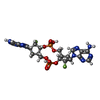  Mass: 658.448 Da / Num. of mol.: 1 / Source method: obtained synthetically / Formula: C22H26F2N10O8P2 / Feature type: SUBJECT OF INVESTIGATION Mass: 658.448 Da / Num. of mol.: 1 / Source method: obtained synthetically / Formula: C22H26F2N10O8P2 / Feature type: SUBJECT OF INVESTIGATION#4: Water | ChemComp-HOH / |  Mass: 18.015 Da / Num. of mol.: 162 / Source method: isolated from a natural source / Formula: H2O Mass: 18.015 Da / Num. of mol.: 162 / Source method: isolated from a natural source / Formula: H2OHas ligand of interest | Y | |
|---|
-Experimental details
-Experiment
| Experiment | Method: X-RAY DIFFRACTION / Number of used crystals: 1 |
|---|
- Sample preparation
| Crystal | Density Matthews: 2.21 Å3/Da / Density % sol: 44.31 % |
|---|---|
| Crystal grow | Temperature: 291 K / Method: vapor diffusion, sitting drop / Details: 0.2 M Lithium acetate, 20% (w/v) PEG 3350 |
-Data collection
| Diffraction | Mean temperature: 100 K / Serial crystal experiment: N |
|---|---|
| Diffraction source | Source: SYNCHROTRON / Site: BESSY  / Beamline: 14.1 / Wavelength: 0.9184 Å / Beamline: 14.1 / Wavelength: 0.9184 Å |
| Detector | Type: DECTRIS PILATUS 6M / Detector: PIXEL / Date: Jan 24, 2019 |
| Radiation | Protocol: SINGLE WAVELENGTH / Monochromatic (M) / Laue (L): M / Scattering type: x-ray |
| Radiation wavelength | Wavelength: 0.9184 Å / Relative weight: 1 |
| Reflection | Resolution: 2→15.96 Å / Num. obs: 35943 / % possible obs: 99.69 % / Redundancy: 22.7 % / Biso Wilson estimate: 33.8 Å2 / CC1/2: 0.999 / CC star: 1 / Rmerge(I) obs: 0.2265 / Rpim(I) all: 0.04809 / Rrim(I) all: 0.2316 / Net I/σ(I): 14.7 |
| Reflection shell | Resolution: 2→2.071 Å / Redundancy: 22.9 % / Rmerge(I) obs: 3.083 / Mean I/σ(I) obs: 1.16 / Num. unique obs: 3515 / CC1/2: 0.438 / CC star: 0.781 / Rpim(I) all: 0.653 / Rrim(I) all: 3.152 / % possible all: 100 |
- Processing
| Software |
| ||||||||||||||||||||||||||||||||||||||||||||||||||||||||||||||||||||||||||||||||||||||||||||||||||
|---|---|---|---|---|---|---|---|---|---|---|---|---|---|---|---|---|---|---|---|---|---|---|---|---|---|---|---|---|---|---|---|---|---|---|---|---|---|---|---|---|---|---|---|---|---|---|---|---|---|---|---|---|---|---|---|---|---|---|---|---|---|---|---|---|---|---|---|---|---|---|---|---|---|---|---|---|---|---|---|---|---|---|---|---|---|---|---|---|---|---|---|---|---|---|---|---|---|---|---|
| Refinement | Method to determine structure: MOLECULAR REPLACEMENT Starting model: 4ksy Resolution: 2→15.96 Å / SU ML: 0.2546 / Cross valid method: FREE R-VALUE / σ(F): 1.34 / Phase error: 25.4384 Stereochemistry target values: GeoStd + Monomer Library + CDL v1.2
| ||||||||||||||||||||||||||||||||||||||||||||||||||||||||||||||||||||||||||||||||||||||||||||||||||
| Solvent computation | Shrinkage radii: 0.9 Å / VDW probe radii: 1.1 Å / Solvent model: FLAT BULK SOLVENT MODEL | ||||||||||||||||||||||||||||||||||||||||||||||||||||||||||||||||||||||||||||||||||||||||||||||||||
| Displacement parameters | Biso mean: 47.08 Å2 | ||||||||||||||||||||||||||||||||||||||||||||||||||||||||||||||||||||||||||||||||||||||||||||||||||
| Refinement step | Cycle: LAST / Resolution: 2→15.96 Å
| ||||||||||||||||||||||||||||||||||||||||||||||||||||||||||||||||||||||||||||||||||||||||||||||||||
| Refine LS restraints |
| ||||||||||||||||||||||||||||||||||||||||||||||||||||||||||||||||||||||||||||||||||||||||||||||||||
| LS refinement shell |
|
