 Movie
Movie Controller

[English] 日本語
 Yorodumi
- PDB-7zw7: Crystal structure of Schistosoma mansoni HDAC8 in complex with a ... -
+ Open data

- Basic information
| Entry | Database: PDB / ID: 7zw7 | ||||||
|---|---|---|---|---|---|---|---|
| Title | Crystal structure of Schistosoma mansoni HDAC8 in complex with a formate molecule in the active site | ||||||
 Components Components | Putative histone deacetylase 1, 2,3 | ||||||
 Keywords Keywords | HYDROLASE / Histone deacetylase 8 / Schistosoma mansoni / Formate | ||||||
| Function / homology |  Function and homology information Function and homology informationhistone deacetylase / histone deacetylase activity / negative regulation of transcription by RNA polymerase II / metal ion binding / nucleus Similarity search - Function | ||||||
| Biological species |  | ||||||
| Method |  X-RAY DIFFRACTION / SYNCHROTRON / MOLECULAR REPLACEMENT / molecular replacement / Resolution: 2.85 Å X-RAY DIFFRACTION / SYNCHROTRON / MOLECULAR REPLACEMENT / molecular replacement / Resolution: 2.85 Å | ||||||
 Authors Authors | Saccoccia, F. / Giannaccari, M. / Ruberti, G. | ||||||
| Funding support |  Italy, 1items Italy, 1items
| ||||||
 Citation Citation | Journal: To Be Published Title: Crystal structure of Schistosoma mansoni HDAC8 in complex with a formate molecule in the active site Authors: Saccoccia, F. / Giannaccari, M. / Ruberti, G. | ||||||
| History |
|
- Structure visualization
| Structure viewer | Molecule: MolmilJmol/JSmol |
|---|
- Downloads & links
-Download
| PDBx/mmCIF format | 7zw7.cif.gz | 180 KB | Display | PDBx/mmCIF format |
|---|---|---|---|---|
| PDB format | pdb7zw7.ent.gz | 141.6 KB | Display | PDB format |
| PDBx/mmJSON format | 7zw7.json.gz | Tree view | PDBx/mmJSON format | |
| Others |  Other downloads Other downloads |
-Validation report
| Summary document | 7zw7_validation.pdf.gz | 885.9 KB | Display | wwPDB validaton report |
|---|---|---|---|---|
| Full document | 7zw7_full_validation.pdf.gz | 894.7 KB | Display | |
| Data in XML | 7zw7_validation.xml.gz | 17.1 KB | Display | |
| Data in CIF | 7zw7_validation.cif.gz | 22.5 KB | Display | |
| Arichive directory | https://data.pdbj.org/pub/pdb/validation_reports/zw/7zw7ftp://data.pdbj.org/pub/pdb/validation_reports/zw/7zw7 | HTTPS FTP |
-Related structure data
| Related structure data | 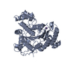 4bz5S S: Starting model for refinement |
|---|---|
| Similar structure data |
-Links
 PDBj
PDBj- Assembly
| Deposited unit | 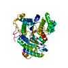
| ||||||||
|---|---|---|---|---|---|---|---|---|---|
| 1 |
| ||||||||
| Unit cell |
|
-Components
-Protein , 1 types, 1 molecules A
| #1: Protein | Mass: 49834.152 Da / Num. of mol.: 1 Source method: isolated from a genetically manipulated source Source: (gene. exp.)  |
|---|
-Non-polymers , 6 types, 16 molecules 


| #2: Chemical | ChemComp-ZN /  Mass: 65.409 Da / Num. of mol.: 1 / Source method: obtained synthetically / Formula: Zn Mass: 65.409 Da / Num. of mol.: 1 / Source method: obtained synthetically / Formula: Zn | ||||||||
|---|---|---|---|---|---|---|---|---|---|
| #3: Chemical |  Mass: 39.098 Da / Num. of mol.: 2 / Source method: obtained synthetically / Formula: K Mass: 39.098 Da / Num. of mol.: 2 / Source method: obtained synthetically / Formula: K#4: Chemical | ChemComp-CL / |  Mass: 35.453 Da / Num. of mol.: 1 / Source method: obtained synthetically / Formula: Cl Mass: 35.453 Da / Num. of mol.: 1 / Source method: obtained synthetically / Formula: Cl#5: Chemical |  Mass: 46.025 Da / Num. of mol.: 2 / Source method: obtained synthetically / Formula: CH2O2 / Feature type: SUBJECT OF INVESTIGATION Mass: 46.025 Da / Num. of mol.: 2 / Source method: obtained synthetically / Formula: CH2O2 / Feature type: SUBJECT OF INVESTIGATION#6: Chemical | ChemComp-PEG / |  Mass: 106.120 Da / Num. of mol.: 1 / Source method: obtained synthetically / Formula: C4H10O3 Mass: 106.120 Da / Num. of mol.: 1 / Source method: obtained synthetically / Formula: C4H10O3#7: Water | ChemComp-HOH / | Mass: 18.015 Da / Num. of mol.: 9 / Source method: isolated from a natural source / Formula: H2O |
-Details
| Has ligand of interest | Y |
|---|
-Experimental details
-Experiment
| Experiment | Method: X-RAY DIFFRACTION / Number of used crystals: 1 |
|---|
- Sample preparation
| Crystal | Density Matthews: 2.27 Å3/Da / Density % sol: 45.9 % |
|---|---|
| Crystal grow | Temperature: 298.15 K / Method: vapor diffusion, hanging drop / pH: 6.5 Details: 20% PEG 3350, 0.2M sodium formate, 0.1M bis-tris propane pH 6.5 |
-Data collection
| Diffraction | Mean temperature: 100 K / Serial crystal experiment: N | ||||||||||||||||||||||||||||||
|---|---|---|---|---|---|---|---|---|---|---|---|---|---|---|---|---|---|---|---|---|---|---|---|---|---|---|---|---|---|---|---|
| Diffraction source | Source: SYNCHROTRON / Site: ELETTRA / Beamline: 11.2C / Wavelength: 1 Å | ||||||||||||||||||||||||||||||
| Detector | Type: DECTRIS PILATUS 6M / Detector: PIXEL / Date: Dec 7, 2021 | ||||||||||||||||||||||||||||||
| Radiation | Protocol: SINGLE WAVELENGTH / Monochromatic (M) / Laue (L): M / Scattering type: x-ray | ||||||||||||||||||||||||||||||
| Radiation wavelength | Wavelength: 1 Å / Relative weight: 1 | ||||||||||||||||||||||||||||||
| Reflection | Resolution: 2.85→45.89 Å / Num. obs: 11416 / % possible obs: 99.9 % / Redundancy: 8.1 % / Biso Wilson estimate: 45.91 Å2 / CC1/2: 0.973 / Rmerge(I) obs: 0.466 / Rpim(I) all: 0.171 / Rrim(I) all: 0.498 / Χ2: 0.99 / Net I/σ(I): 4.6 | ||||||||||||||||||||||||||||||
| Reflection shell | Diffraction-ID: 1
|
-Phasing
| Phasing | Method: molecular replacement | |||||||||
|---|---|---|---|---|---|---|---|---|---|---|
| Phasing MR | Model details: Phaser MODE: MR_AUTO
|
- Processing
| Software |
| ||||||||||||||||||||||||||||||||||||||||||||||||||||||||
|---|---|---|---|---|---|---|---|---|---|---|---|---|---|---|---|---|---|---|---|---|---|---|---|---|---|---|---|---|---|---|---|---|---|---|---|---|---|---|---|---|---|---|---|---|---|---|---|---|---|---|---|---|---|---|---|---|---|
| Refinement | Method to determine structure: MOLECULAR REPLACEMENT Starting model: 4BZ5 Resolution: 2.85→45.89 Å / SU ML: 0.38 / Cross valid method: THROUGHOUT / σ(F): 0.2 / Phase error: 26.97 / Stereochemistry target values: ML
| ||||||||||||||||||||||||||||||||||||||||||||||||||||||||
| Solvent computation | Shrinkage radii: 0.8 Å / VDW probe radii: 1 Å / Solvent model: FLAT BULK SOLVENT MODEL | ||||||||||||||||||||||||||||||||||||||||||||||||||||||||
| Displacement parameters | Biso max: 88.51 Å2 / Biso mean: 46.1877 Å2 / Biso min: 32.08 Å2 | ||||||||||||||||||||||||||||||||||||||||||||||||||||||||
| Refinement step | Cycle: final / Resolution: 2.85→45.89 Å
| ||||||||||||||||||||||||||||||||||||||||||||||||||||||||
| LS refinement shell | Refine-ID: X-RAY DIFFRACTION / Rfactor Rfree error: 0 / Total num. of bins used: 7
| ||||||||||||||||||||||||||||||||||||||||||||||||||||||||
| Refinement TLS params. | Method: refined / Origin x: -8.614 Å / Origin y: 22.0787 Å / Origin z: 15.4252 Å
| ||||||||||||||||||||||||||||||||||||||||||||||||||||||||
| Refinement TLS group |
|
