 Movie
Movie Controller

[English] 日本語
 Yorodumi
- PDB-7zvp: Crystal structure of poplar glutathione transferase U19 in comple... -
+ Open data

- Basic information
| Entry | Database: PDB / ID: 7zvp | ||||||
|---|---|---|---|---|---|---|---|
| Title | Crystal structure of poplar glutathione transferase U19 in complex with glutathione | ||||||
 Components Components | Glutathione transferase | ||||||
 Keywords Keywords | TRANSFERASE / Glutathione / Tau | ||||||
| Function / homology |  Function and homology information Function and homology informationglutathione transferase / glutathione transferase activity / glutathione metabolic process / cytoplasm / cytosol Similarity search - Function | ||||||
| Biological species |  | ||||||
| Method |  X-RAY DIFFRACTION / SYNCHROTRON / MOLECULAR REPLACEMENT / Resolution: 1.61 Å X-RAY DIFFRACTION / SYNCHROTRON / MOLECULAR REPLACEMENT / Resolution: 1.61 Å | ||||||
 Authors Authors | Didierjean, C. / Favier, F. | ||||||
| Funding support |  France, 1items France, 1items
| ||||||
 Citation Citation | Journal: Front Mol Biosci / Year: 2022 Title: Biochemical and Structural Insights on the Poplar Tau Glutathione Transferase GSTU19 and 20 Paralogs Binding Flavonoids. Authors: Sylvestre-Gonon, E. / Morette, L. / Viloria, M. / Mathiot, S. / Boutilliat, A. / Favier, F. / Rouhier, N. / Didierjean, C. / Hecker, A. | ||||||
| History |
|
- Structure visualization
| Structure viewer | Molecule: MolmilJmol/JSmol |
|---|
- Downloads & links
-Download
| PDBx/mmCIF format | 7zvp.cif.gz | 65.5 KB | Display | PDBx/mmCIF format |
|---|---|---|---|---|
| PDB format | pdb7zvp.ent.gz | 46 KB | Display | PDB format |
| PDBx/mmJSON format | 7zvp.json.gz | Tree view | PDBx/mmJSON format | |
| Others |  Other downloads Other downloads |
-Validation report
| Arichive directory | https://data.pdbj.org/pub/pdb/validation_reports/zv/7zvpftp://data.pdbj.org/pub/pdb/validation_reports/zv/7zvp | HTTPS FTP |
|---|
-Related structure data
| Related structure data |  7zs3SC  7zznC 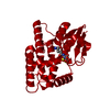 8a08C  8a0iC  8a0oC  8a0pC 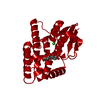 8a0qC  8a0rC S: Starting model for refinement C: citing same article ( |
|---|---|
| Similar structure data |
-Links
 PDBj
PDBj

- Assembly
| Deposited unit | 
| ||||||||
|---|---|---|---|---|---|---|---|---|---|
| 1 | 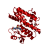
| ||||||||
| Unit cell |
| ||||||||
| Components on special symmetry positions |
|
-Components
| #1: Protein | Mass: 25053.969 Da / Num. of mol.: 1 Source method: isolated from a genetically manipulated source Source: (gene. exp.)  | ||||
|---|---|---|---|---|---|
| #2: Chemical | ChemComp-GS8 / 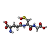  Mass: 323.323 Da / Num. of mol.: 1 / Source method: obtained synthetically / Formula: C10H17N3O7S / Feature type: SUBJECT OF INVESTIGATION Mass: 323.323 Da / Num. of mol.: 1 / Source method: obtained synthetically / Formula: C10H17N3O7S / Feature type: SUBJECT OF INVESTIGATION | ||||
| #3: Chemical |   Mass: 59.044 Da / Num. of mol.: 2 / Source method: obtained synthetically / Formula: C2H3O2 Mass: 59.044 Da / Num. of mol.: 2 / Source method: obtained synthetically / Formula: C2H3O2#4: Water | ChemComp-HOH / |  Mass: 18.015 Da / Num. of mol.: 257 / Source method: isolated from a natural source / Formula: H2O Mass: 18.015 Da / Num. of mol.: 257 / Source method: isolated from a natural source / Formula: H2OHas ligand of interest | Y | |
-Experimental details
-Experiment
| Experiment | Method: X-RAY DIFFRACTION / Number of used crystals: 1 |
|---|
- Sample preparation
| Crystal | Density Matthews: 2.92 Å3/Da / Density % sol: 57.91 % |
|---|---|
| Crystal grow | Temperature: 277 K / Method: microbatch / pH: 6.5 Details: 0.2 M Calcium acetate hydrate, 0.1 M Sodium cacodylate pH 6.5 and 18 % w/v PEG 8000 |
-Data collection
| Diffraction | Mean temperature: 100 K / Serial crystal experiment: N |
|---|---|
| Diffraction source | Source: SYNCHROTRON / Site: SOLEIL / Beamline: PROXIMA 2 / Wavelength: 0.9801 Å |
| Detector | Type: DECTRIS EIGER X 9M / Detector: PIXEL / Date: Jun 16, 2019 |
| Radiation | Protocol: SINGLE WAVELENGTH / Monochromatic (M) / Laue (L): M / Scattering type: x-ray |
| Radiation wavelength | Wavelength: 0.9801 Å / Relative weight: 1 |
| Reflection | Resolution: 1.61→48.1 Å / Num. obs: 39646 / % possible obs: 99.9 % / Redundancy: 10 % / Biso Wilson estimate: 26.7 Å2 / CC1/2: 1 / Rmerge(I) obs: 0.062 / Net I/σ(I): 17.7 |
| Reflection shell | Resolution: 1.61→1.64 Å / Redundancy: 10 % / Rmerge(I) obs: 0.67 / Mean I/σ(I) obs: 2.3 / Num. unique obs: 1927 / CC1/2: 0.94 / % possible all: 100 |
- Processing
| Software |
| ||||||||||||||||||||||||||||||||||||||||||||||||||||||||||||||||||
|---|---|---|---|---|---|---|---|---|---|---|---|---|---|---|---|---|---|---|---|---|---|---|---|---|---|---|---|---|---|---|---|---|---|---|---|---|---|---|---|---|---|---|---|---|---|---|---|---|---|---|---|---|---|---|---|---|---|---|---|---|---|---|---|---|---|---|---|
| Refinement | Method to determine structure: MOLECULAR REPLACEMENT Starting model: 7ZS3 Resolution: 1.61→48.14 Å / Cor.coef. Fo:Fc: 0.948 / Cor.coef. Fo:Fc free: 0.942 / SU R Cruickshank DPI: 0.095 / Cross valid method: THROUGHOUT / SU R Blow DPI: 0.103 / SU Rfree Blow DPI: 0.098 / SU Rfree Cruickshank DPI: 0.092
| ||||||||||||||||||||||||||||||||||||||||||||||||||||||||||||||||||
| Displacement parameters | Biso mean: 32.07 Å2
| ||||||||||||||||||||||||||||||||||||||||||||||||||||||||||||||||||
| Refine analyze | Luzzati coordinate error obs: 0.27 Å | ||||||||||||||||||||||||||||||||||||||||||||||||||||||||||||||||||
| Refinement step | Cycle: LAST / Resolution: 1.61→48.14 Å
| ||||||||||||||||||||||||||||||||||||||||||||||||||||||||||||||||||
| Refine LS restraints |
| ||||||||||||||||||||||||||||||||||||||||||||||||||||||||||||||||||
| LS refinement shell | Resolution: 1.61→1.62 Å
|
