| Entry | Database: PDB / ID: 7zvd
|
|---|
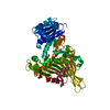
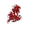
| Title | K89 acetylated glucose-6-phosphate dehydrogenase (G6PD) in a complex with structural NADP+ |
|---|
 Components Components | Glucose-6-phosphate 1-dehydrogenase |
|---|
 Keywords Keywords | OXIDOREDUCTASE / Lysine acetylation / metabolic regulation / post-translational modification / genetic code expansion |
|---|
| Function / homology |  Function and homology information Function and homology information
pentose biosynthetic process / ribose phosphate biosynthetic process / response to iron(III) ion / positive regulation of calcium ion transmembrane transport via high voltage-gated calcium channel / glucose-6-phosphate dehydrogenase (NADP+) / glucose-6-phosphate dehydrogenase activity / Pentose phosphate pathway / pentose-phosphate shunt, oxidative branch / negative regulation of cell growth involved in cardiac muscle cell development / NADPH regeneration ...pentose biosynthetic process / ribose phosphate biosynthetic process / response to iron(III) ion / positive regulation of calcium ion transmembrane transport via high voltage-gated calcium channel / glucose-6-phosphate dehydrogenase (NADP+) / glucose-6-phosphate dehydrogenase activity / Pentose phosphate pathway / pentose-phosphate shunt, oxidative branch / negative regulation of cell growth involved in cardiac muscle cell development / NADPH regeneration / glucose 6-phosphate metabolic process / NADP+ metabolic process / pentose-phosphate shunt / D-glucose binding / NFE2L2 regulates pentose phosphate pathway genes / response to food / erythrocyte maturation / cholesterol biosynthetic process / negative regulation of reactive oxygen species metabolic process / regulation of neuron apoptotic process / glutathione metabolic process / substantia nigra development / TP53 Regulates Metabolic Genes / lipid metabolic process / centriolar satellite / cytoplasmic side of plasma membrane / glucose metabolic process / NADP binding / cellular response to oxidative stress / response to ethanol / intracellular membrane-bounded organelle / protein homodimerization activity / extracellular exosome / identical protein binding / membrane / cytoplasm / cytosolSimilarity search - Function Glucose-6-phosphate dehydrogenase, active site / Glucose-6-phosphate dehydrogenase active site. / Glucose-6-phosphate dehydrogenase / Glucose-6-phosphate dehydrogenase, NAD-binding / Glucose-6-phosphate dehydrogenase, C-terminal / Glucose-6-phosphate dehydrogenase, NAD binding domain / Glucose-6-phosphate dehydrogenase, C-terminal domain / NAD(P)-binding domain superfamilySimilarity search - Domain/homology |
|---|
| Biological species |  Homo sapiens (human) Homo sapiens (human) |
|---|
| Method |  X-RAY DIFFRACTION / SYNCHROTRON / MOLECULAR REPLACEMENT / Resolution: 2.46 Å X-RAY DIFFRACTION / SYNCHROTRON / MOLECULAR REPLACEMENT / Resolution: 2.46 Å |
|---|
 Authors Authors | Wu, F. / Muskat, N.H. / Nudelman, H. / Shahar, A. / Arbely, E. |
|---|
| Funding support | European Union,  Israel, 2items Israel, 2items | Organization | Grant number | Country |
|---|
| European Research Council (ERC) | 678461 | European Union | | Israel Science Foundation | 1769/20 | Israel |
|
|---|
 Citation Citation | Journal: Nat Commun / Year: 2023
Title: Acetylation-dependent coupling between G6PD activity and apoptotic signaling.
Authors: Wu, F. / Muskat, N.H. / Dvilansky, I. / Koren, O. / Shahar, A. / Gazit, R. / Elia, N. / Arbely, E. |
|---|
| History | | Deposition | May 15, 2022 | Deposition site: PDBE / Processing site: PDBE |
|---|
| Revision 1.0 | Aug 23, 2023 | Provider: repository / Type: Initial release |
|---|
| Revision 1.1 | Nov 15, 2023 | Group: Data collection / Category: chem_comp_atom / chem_comp_bond / Item: _chem_comp_atom.atom_id / _chem_comp_bond.atom_id_2 |
|---|
| Revision 1.2 | Feb 7, 2024 | Group: Database references / Category: citation / citation_author
Item: _citation.country / _citation.journal_abbrev ..._citation.country / _citation.journal_abbrev / _citation.journal_id_CSD / _citation.journal_id_ISSN / _citation.journal_volume / _citation.page_first / _citation.page_last / _citation.pdbx_database_id_DOI / _citation.pdbx_database_id_PubMed / _citation.title / _citation.year |
|---|
| Revision 1.3 | Feb 28, 2024 | Group: Source and taxonomy / Category: entity_src_gen
Item: _entity_src_gen.pdbx_gene_src_ncbi_taxonomy_id / _entity_src_gen.pdbx_gene_src_scientific_name |
|---|
| Revision 1.4 | Nov 13, 2024 | Group: Structure summary / Category: pdbx_entry_details / pdbx_modification_feature / Item: _pdbx_entry_details.has_protein_modification |
|---|
|
|---|
 Movie
Movie Controller

 Yorodumi
 Open data
 Basic information
 Structure visualization
 Downloads & links
 Other downloads
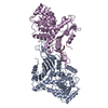
 PDBj
PDBj Assembly
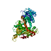


 Mass: 743.405 Da / Num. of mol.: 1 / Source method: obtained synthetically / Formula: C21H28N7O17P3 / Feature type: SUBJECT OF INVESTIGATION
 Mass: 18.015 Da / Num. of mol.: 46 / Source method: isolated from a natural source / Formula: H2O
 Sample preparation
 / Beamline: I03 / Wavelength: 0.9763 Å
 Processing