 Movie
Movie Controller


+ Open data

- Basic information
| Entry | Database: PDB / ID: 7zoz | |||||||||
|---|---|---|---|---|---|---|---|---|---|---|
| Title | Crystal structure of Siglec-15 in complex with Fab | |||||||||
 Components Components |
| |||||||||
 Keywords Keywords | IMMUNE SYSTEM / sialic acid / siglec / cancer | |||||||||
| Function / homology |  Function and homology information Function and homology informationregulation of osteoclast development / regulation of bone resorption / DAP12 interactions / regulation of actin cytoskeleton organization / protein-containing complex / plasma membrane Similarity search - Function | |||||||||
| Biological species |  Homo sapiens (human) Homo sapiens (human) | |||||||||
| Method |  X-RAY DIFFRACTION / SYNCHROTRON / MOLECULAR REPLACEMENT / Resolution: 2.104 Å X-RAY DIFFRACTION / SYNCHROTRON / MOLECULAR REPLACEMENT / Resolution: 2.104 Å | |||||||||
 Authors Authors | Lenza, M.P. / Oyenarte, I. / Jimenez-Barbero, J. / Ereno-Orbea, J. | |||||||||
| Funding support | European Union,  Spain, 2items Spain, 2items
| |||||||||
 Citation Citation | Journal: Nat Commun / Year: 2023 Title: Structural insights into Siglec-15 reveal glycosylation dependency for its interaction with T cells through integrin CD11b. Authors: Lenza, M.P. / Egia-Mendikute, L. / Antonana-Vildosola, A. / Soares, C.O. / Coelho, H. / Corzana, F. / Bosch, A. / Manisha, P. / Quintana, J.I. / Oyenarte, I. / Unione, L. / Moure, M.J. / ...Authors: Lenza, M.P. / Egia-Mendikute, L. / Antonana-Vildosola, A. / Soares, C.O. / Coelho, H. / Corzana, F. / Bosch, A. / Manisha, P. / Quintana, J.I. / Oyenarte, I. / Unione, L. / Moure, M.J. / Azkargorta, M. / Atxabal, U. / Sobczak, K. / Elortza, F. / Sutherland, J.D. / Barrio, R. / Marcelo, F. / Jimenez-Barbero, J. / Palazon, A. / Ereno-Orbea, J. | |||||||||
| History |
|
- Structure visualization
| Structure viewer | Molecule: MolmilJmol/JSmol |
|---|
- Downloads & links
-Download
| PDBx/mmCIF format | 7zoz.cif.gz | 249.1 KB | Display | PDBx/mmCIF format |
|---|---|---|---|---|
| PDB format | pdb7zoz.ent.gz | 194.7 KB | Display | PDB format |
| PDBx/mmJSON format | 7zoz.json.gz | Tree view | PDBx/mmJSON format | |
| Others |  Other downloads Other downloads |
-Validation report
| Summary document | 7zoz_validation.pdf.gz | 435.5 KB | Display | wwPDB validaton report |
|---|---|---|---|---|
| Full document | 7zoz_full_validation.pdf.gz | 436.6 KB | Display | |
| Data in XML | 7zoz_validation.xml.gz | 23.9 KB | Display | |
| Data in CIF | 7zoz_validation.cif.gz | 34.9 KB | Display | |
| Arichive directory | https://data.pdbj.org/pub/pdb/validation_reports/zo/7zozftp://data.pdbj.org/pub/pdb/validation_reports/zo/7zoz | HTTPS FTP |
-Related structure data
| Related structure data | 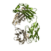 7zorC 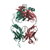 5vkkS C: citing same article ( S: Starting model for refinement |
|---|---|
| Similar structure data |
-Links
 PDBj
PDBj


- Assembly
| Deposited unit | 
| ||||||||
|---|---|---|---|---|---|---|---|---|---|
| 1 |
| ||||||||
| Unit cell |
|
-Components
| #1: Protein | Mass: 33598.988 Da / Num. of mol.: 1 Source method: isolated from a genetically manipulated source Source: (gene. exp.) Homo sapiens (human) / Gene: SIGLEC15, CD33L3 / Production host: Homo sapiens (human) / References: UniProt: Q6ZMC9 |
|---|---|
| #2: Antibody | Mass: 24378.148 Da / Num. of mol.: 1 Source method: isolated from a genetically manipulated source Source: (gene. exp.) Homo sapiens (human) / Production host: Homo sapiens (human) |
| #3: Antibody | Mass: 23765.467 Da / Num. of mol.: 1 Source method: isolated from a genetically manipulated source Source: (gene. exp.) Homo sapiens (human) / Production host: Homo sapiens (human) |
| #4: Water | ChemComp-HOH /  Mass: 18.015 Da / Num. of mol.: 305 / Source method: isolated from a natural source / Formula: H2O Mass: 18.015 Da / Num. of mol.: 305 / Source method: isolated from a natural source / Formula: H2O |
| Has protein modification | Y |
-Experimental details
-Experiment
| Experiment | Method: X-RAY DIFFRACTION / Number of used crystals: 1 |
|---|
- Sample preparation
| Crystal | Density Matthews: 2.05 Å3/Da / Density % sol: 39.96 % |
|---|---|
| Crystal grow | Temperature: 291 K / Method: vapor diffusion, hanging drop / Details: 20 % PEG 3350 0.2 M Calcium Chloride |
-Data collection
| Diffraction | Mean temperature: 100 K / Serial crystal experiment: N |
|---|---|
| Diffraction source | Source: SYNCHROTRON / Site: SLS  / Beamline: X06DA / Wavelength: 0.97925 Å / Beamline: X06DA / Wavelength: 0.97925 Å |
| Detector | Type: DECTRIS PILATUS 2M-F / Detector: PIXEL / Date: Sep 11, 2020 |
| Radiation | Protocol: SINGLE WAVELENGTH / Monochromatic (M) / Laue (L): M / Scattering type: x-ray |
| Radiation wavelength | Wavelength: 0.97925 Å / Relative weight: 1 |
| Reflection | Resolution: 2.104→106.342 Å / Num. obs: 37172 / % possible obs: 99.8 % / Redundancy: 6.5 % / Biso Wilson estimate: 33.79 Å2 / CC1/2: 0.998 / Rmerge(I) obs: 0.078 / Rpim(I) all: 0.033 / Rrim(I) all: 0.084 / Net I/σ(I): 15.1 |
| Reflection shell | Resolution: 2.104→2.18 Å / Rmerge(I) obs: 0.633 / Num. unique obs: 1824 / CC1/2: 0.835 / Rpim(I) all: 0.307 / Rrim(I) all: 0.707 |
- Processing
| Software |
| ||||||||||||||||||||||||||||||||||||||||||||||||||||||||||||||||||||||||||||||||||||||||||
|---|---|---|---|---|---|---|---|---|---|---|---|---|---|---|---|---|---|---|---|---|---|---|---|---|---|---|---|---|---|---|---|---|---|---|---|---|---|---|---|---|---|---|---|---|---|---|---|---|---|---|---|---|---|---|---|---|---|---|---|---|---|---|---|---|---|---|---|---|---|---|---|---|---|---|---|---|---|---|---|---|---|---|---|---|---|---|---|---|---|---|---|
| Refinement | Method to determine structure: MOLECULAR REPLACEMENT Starting model: 5vkk Resolution: 2.104→106.341 Å / SU ML: 0.24 / Cross valid method: THROUGHOUT / σ(F): 1.35 / Phase error: 23.34 / Stereochemistry target values: ML
| ||||||||||||||||||||||||||||||||||||||||||||||||||||||||||||||||||||||||||||||||||||||||||
| Solvent computation | Shrinkage radii: 0.9 Å / VDW probe radii: 1.11 Å / Solvent model: FLAT BULK SOLVENT MODEL | ||||||||||||||||||||||||||||||||||||||||||||||||||||||||||||||||||||||||||||||||||||||||||
| Displacement parameters | Biso max: 127.58 Å2 / Biso mean: 40.4562 Å2 / Biso min: 17 Å2 | ||||||||||||||||||||||||||||||||||||||||||||||||||||||||||||||||||||||||||||||||||||||||||
| Refinement step | Cycle: final / Resolution: 2.104→106.341 Å
| ||||||||||||||||||||||||||||||||||||||||||||||||||||||||||||||||||||||||||||||||||||||||||
| LS refinement shell | Refine-ID: X-RAY DIFFRACTION / Rfactor Rfree error: 0
| ||||||||||||||||||||||||||||||||||||||||||||||||||||||||||||||||||||||||||||||||||||||||||
| Refinement TLS params. | Method: refined / Origin x: -37.9702 Å / Origin y: 1.4837 Å / Origin z: 14.5035 Å
| ||||||||||||||||||||||||||||||||||||||||||||||||||||||||||||||||||||||||||||||||||||||||||
| Refinement TLS group |
|
