 Movie
Movie Controller

[English] 日本語
 Yorodumi
- PDB-7zoy: Crystal structure of Synechocystis halorhodopsin (SyHR), SO4-boun... -
+ Open data

- Basic information
| Entry | Database: PDB / ID: 7zoy | ||||||||||||||||||
|---|---|---|---|---|---|---|---|---|---|---|---|---|---|---|---|---|---|---|---|
| Title | Crystal structure of Synechocystis halorhodopsin (SyHR), SO4-bound form, ground state | ||||||||||||||||||
 Components Components | Synechocystis halorhodopsin | ||||||||||||||||||
 Keywords Keywords | MEMBRANE PROTEIN / rhodopsin / syhr / trapping / cryo / cryotrapping / o-state / k-state / sulfate / chloride / pump / anion / photocycle / ion transport | ||||||||||||||||||
| Function / homology | EICOSANE / OLEIC ACID Function and homology information Function and homology information | ||||||||||||||||||
| Biological species |  | ||||||||||||||||||
| Method |  X-RAY DIFFRACTION / SYNCHROTRON / MOLECULAR REPLACEMENT / Resolution: 1.91 Å X-RAY DIFFRACTION / SYNCHROTRON / MOLECULAR REPLACEMENT / Resolution: 1.91 Å | ||||||||||||||||||
 Authors Authors | Kovalev, K. / Bukhdruker, S. / Astashkin, R. / Vaganova, S. / Gordeliy, V. | ||||||||||||||||||
| Funding support |  France, France,  Russian Federation, 5items Russian Federation, 5items
| ||||||||||||||||||
 Citation Citation | Journal: Nat Commun / Year: 2022 Title: Structural insights into light-driven anion pumping in cyanobacteria. Authors: Astashkin, R. / Kovalev, K. / Bukhdruker, S. / Vaganova, S. / Kuzmin, A. / Alekseev, A. / Balandin, T. / Zabelskii, D. / Gushchin, I. / Royant, A. / Volkov, D. / Bourenkov, G. / Koonin, E. / ...Authors: Astashkin, R. / Kovalev, K. / Bukhdruker, S. / Vaganova, S. / Kuzmin, A. / Alekseev, A. / Balandin, T. / Zabelskii, D. / Gushchin, I. / Royant, A. / Volkov, D. / Bourenkov, G. / Koonin, E. / Engelhard, M. / Bamberg, E. / Gordeliy, V. | ||||||||||||||||||
| History |
|
- Structure visualization
| Structure viewer | Molecule: MolmilJmol/JSmol |
|---|
- Downloads & links
-Download
| PDBx/mmCIF format | 7zoy.cif.gz | 78.1 KB | Display | PDBx/mmCIF format |
|---|---|---|---|---|
| PDB format | pdb7zoy.ent.gz | 49.7 KB | Display | PDB format |
| PDBx/mmJSON format | 7zoy.json.gz | Tree view | PDBx/mmJSON format | |
| Others |  Other downloads Other downloads |
-Validation report
| Summary document | 7zoy_validation.pdf.gz | 2.9 MB | Display | wwPDB validaton report |
|---|---|---|---|---|
| Full document | 7zoy_full_validation.pdf.gz | 2.9 MB | Display | |
| Data in XML | 7zoy_validation.xml.gz | 13.4 KB | Display | |
| Data in CIF | 7zoy_validation.cif.gz | 17.6 KB | Display | |
| Arichive directory | https://data.pdbj.org/pub/pdb/validation_reports/zo/7zoyftp://data.pdbj.org/pub/pdb/validation_reports/zo/7zoy | HTTPS FTP |
-Related structure data
| Related structure data |  7zouC 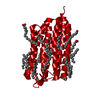 7zovC  7zowC 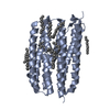 4hyjS S: Starting model for refinement C: citing same article ( |
|---|---|
| Similar structure data |
-Links
 PDBj
PDBj


- Assembly
| Deposited unit | 
| ||||||||||||
|---|---|---|---|---|---|---|---|---|---|---|---|---|---|
| 1 | 
| ||||||||||||
| Unit cell |
| ||||||||||||
| Components on special symmetry positions |
|
-Components
-Protein , 1 types, 1 molecules A
| #1: Protein | Mass: 26463.045 Da / Num. of mol.: 1 Source method: isolated from a genetically manipulated source Source: (gene. exp.) |
|---|
-Non-polymers , 6 types, 92 molecules 


| #2: Chemical | ChemComp-CL /  Mass: 35.453 Da / Num. of mol.: 1 / Source method: obtained synthetically / Formula: Cl Mass: 35.453 Da / Num. of mol.: 1 / Source method: obtained synthetically / Formula: Cl | ||||||
|---|---|---|---|---|---|---|---|
| #3: Chemical | ChemComp-SO4 /  Mass: 96.063 Da / Num. of mol.: 1 / Source method: obtained synthetically / Formula: SO4 / Feature type: SUBJECT OF INVESTIGATION Mass: 96.063 Da / Num. of mol.: 1 / Source method: obtained synthetically / Formula: SO4 / Feature type: SUBJECT OF INVESTIGATION | ||||||
| #4: Chemical | ChemComp-LFA /  Mass: 282.547 Da / Num. of mol.: 13 / Source method: obtained synthetically / Formula: C20H42 Mass: 282.547 Da / Num. of mol.: 13 / Source method: obtained synthetically / Formula: C20H42#5: Chemical |  Mass: 282.461 Da / Num. of mol.: 2 / Source method: obtained synthetically / Formula: C18H34O2 Mass: 282.461 Da / Num. of mol.: 2 / Source method: obtained synthetically / Formula: C18H34O2#6: Chemical | ChemComp-GOL / |  Mass: 92.094 Da / Num. of mol.: 1 / Source method: obtained synthetically / Formula: C3H8O3 Mass: 92.094 Da / Num. of mol.: 1 / Source method: obtained synthetically / Formula: C3H8O3#7: Water | ChemComp-HOH / | Mass: 18.015 Da / Num. of mol.: 74 / Source method: isolated from a natural source / Formula: H2O |
-Details
| Has ligand of interest | Y |
|---|
-Experimental details
-Experiment
| Experiment | Method: X-RAY DIFFRACTION / Number of used crystals: 1 |
|---|
- Sample preparation
| Crystal | Density Matthews: 2.41 Å3/Da / Density % sol: 49.07 % |
|---|---|
| Crystal grow | Temperature: 293 K / Method: lipidic cubic phase / pH: 7 / Details: 2.0 M Ammonium Sulfate, 0.1 M HEPES |
-Data collection
| Diffraction | Mean temperature: 100 K / Serial crystal experiment: N |
|---|---|
| Diffraction source | Source: SYNCHROTRON / Site: SLS  / Beamline: X10SA / Wavelength: 1 Å / Beamline: X10SA / Wavelength: 1 Å |
| Detector | Type: DECTRIS PILATUS 6M / Detector: PIXEL / Date: Feb 24, 2019 |
| Radiation | Protocol: SINGLE WAVELENGTH / Monochromatic (M) / Laue (L): M / Scattering type: x-ray |
| Radiation wavelength | Wavelength: 1 Å / Relative weight: 1 |
| Reflection | Resolution: 1.91→48.45 Å / Num. obs: 19823 / % possible obs: 99.6 % / Redundancy: 10.7 % / Biso Wilson estimate: 25.53 Å2 / CC1/2: 0.997 / Rpim(I) all: 0.081 / Net I/σ(I): 7.5 |
| Reflection shell | Resolution: 1.91→1.94 Å / Mean I/σ(I) obs: 0.9 / Num. unique obs: 992 / CC1/2: 0.399 / Rpim(I) all: 0.816 |
- Processing
| Software |
| ||||||||||||||||||||||||||||||||||||||||||||||||||||||||
|---|---|---|---|---|---|---|---|---|---|---|---|---|---|---|---|---|---|---|---|---|---|---|---|---|---|---|---|---|---|---|---|---|---|---|---|---|---|---|---|---|---|---|---|---|---|---|---|---|---|---|---|---|---|---|---|---|---|
| Refinement | Method to determine structure: MOLECULAR REPLACEMENT Starting model: 4HYJ Resolution: 1.91→19.27 Å / SU ML: 0.2342 / Cross valid method: FREE R-VALUE / σ(F): 1.34 / Phase error: 24.2934 Stereochemistry target values: GeoStd + Monomer Library + CDL v1.2
| ||||||||||||||||||||||||||||||||||||||||||||||||||||||||
| Solvent computation | Shrinkage radii: 0.9 Å / VDW probe radii: 1.11 Å / Solvent model: FLAT BULK SOLVENT MODEL | ||||||||||||||||||||||||||||||||||||||||||||||||||||||||
| Displacement parameters | Biso mean: 26.97 Å2 | ||||||||||||||||||||||||||||||||||||||||||||||||||||||||
| Refinement step | Cycle: LAST / Resolution: 1.91→19.27 Å
| ||||||||||||||||||||||||||||||||||||||||||||||||||||||||
| Refine LS restraints |
| ||||||||||||||||||||||||||||||||||||||||||||||||||||||||
| LS refinement shell |
|
