 Movie
Movie Controller


+ Open data

- Basic information
| Entry | Database: PDB / ID: 7zot | ||||||
|---|---|---|---|---|---|---|---|
| Title | crystal structure of PLAAT4 N-terminal domain | ||||||
 Components Components | Phospholipase A and acyltransferase 4 | ||||||
 Keywords Keywords | HYDROLASE / phospholipase A1/A2 / acyltransferase / catalytic domain / soluble domain / cytosolic protein | ||||||
| Function / homology |  Function and homology information Function and homology informationpositive regulation of protein-glutamine gamma-glutamyltransferase activity / phosphatidylethanolamine acyl-chain remodeling / N-acylphosphatidylethanolamine metabolic process / phospholipase A1 / Acyl chain remodelling of PE / phospholipase A1 activity / N-acyltransferase activity / phospholipase A2 activity / positive regulation of keratinocyte differentiation / phospholipase A2 ...positive regulation of protein-glutamine gamma-glutamyltransferase activity / phosphatidylethanolamine acyl-chain remodeling / N-acylphosphatidylethanolamine metabolic process / phospholipase A1 / Acyl chain remodelling of PE / phospholipase A1 activity / N-acyltransferase activity / phospholipase A2 activity / positive regulation of keratinocyte differentiation / phospholipase A2 / acyltransferase activity / lipid catabolic process / phospholipid metabolic process / Transferases; Acyltransferases; Transferring groups other than aminoacyl groups / negative regulation of cell population proliferation / membrane / cytoplasm / cytosol Similarity search - Function | ||||||
| Biological species |  Homo sapiens (human) Homo sapiens (human) | ||||||
| Method |  X-RAY DIFFRACTION / SYNCHROTRON / MOLECULAR REPLACEMENT / Resolution: 1.735 Å X-RAY DIFFRACTION / SYNCHROTRON / MOLECULAR REPLACEMENT / Resolution: 1.735 Å | ||||||
 Authors Authors | von Castelmur, E. / Perrakis, A. / Cornaciu, I. | ||||||
| Funding support |  Switzerland, 1items Switzerland, 1items
| ||||||
 Citation Citation | Journal: J.Struct.Biol. / Year: 2022 Title: Crystal structure of the phospholipase A and acyltransferase 4 (PLAAT4) catalytic domain. Authors: Wehlin, A. / Cornaciu, I. / Marquez, J.A. / Perrakis, A. / von Castelmur, E. | ||||||
| History |
|
- Structure visualization
| Structure viewer | Molecule: MolmilJmol/JSmol |
|---|
- Downloads & links
-Download
| PDBx/mmCIF format | 7zot.cif.gz | 64 KB | Display | PDBx/mmCIF format |
|---|---|---|---|---|
| PDB format | pdb7zot.ent.gz | 45.1 KB | Display | PDB format |
| PDBx/mmJSON format | 7zot.json.gz | Tree view | PDBx/mmJSON format | |
| Others |  Other downloads Other downloads |
-Validation report
| Summary document | 7zot_validation.pdf.gz | 396.9 KB | Display | wwPDB validaton report |
|---|---|---|---|---|
| Full document | 7zot_full_validation.pdf.gz | 397.1 KB | Display | |
| Data in XML | 7zot_validation.xml.gz | 6.2 KB | Display | |
| Data in CIF | 7zot_validation.cif.gz | 9.5 KB | Display | |
| Arichive directory | https://data.pdbj.org/pub/pdb/validation_reports/zo/7zotftp://data.pdbj.org/pub/pdb/validation_reports/zo/7zot | HTTPS FTP |
-Related structure data
| Related structure data |  7zomS S: Starting model for refinement |
|---|---|
| Similar structure data |
-Links
 PDBj
PDBj

- Assembly
| Deposited unit | 
| ||||||||
|---|---|---|---|---|---|---|---|---|---|
| 1 | 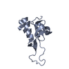
| ||||||||
| 2 | 
| ||||||||
| Unit cell |
|
-Components
| #1: Protein | Mass: 14045.846 Da / Num. of mol.: 2 Source method: isolated from a genetically manipulated source Source: (gene. exp.) Homo sapiens (human) / Gene: PLAAT4, RARRES3, RIG1, TIG3 / Production host:  References: UniProt: Q9UL19, Transferases; Acyltransferases; Transferring groups other than aminoacyl groups, phospholipase A1, phospholipase A2 #2: Chemical | ChemComp-PEG / |   Mass: 106.120 Da / Num. of mol.: 1 / Source method: obtained synthetically / Formula: C4H10O3 Mass: 106.120 Da / Num. of mol.: 1 / Source method: obtained synthetically / Formula: C4H10O3#3: Water | ChemComp-HOH / |  Mass: 18.015 Da / Num. of mol.: 98 / Source method: isolated from a natural source / Formula: H2O Mass: 18.015 Da / Num. of mol.: 98 / Source method: isolated from a natural source / Formula: H2OHas ligand of interest | N | |
|---|
-Experimental details
-Experiment
| Experiment | Method: X-RAY DIFFRACTION / Number of used crystals: 1 |
|---|
- Sample preparation
| Crystal | Density Matthews: 2.17 Å3/Da / Density % sol: 43.27 % |
|---|---|
| Crystal grow | Temperature: 293 K / Method: vapor diffusion, sitting drop / pH: 8.5 Details: Morpheus screen condition H12 0.1M Amino acids, 0.1M Tris Bicine pH 8.5 37.5% v/v MPD PEG1000 PEG 3350 |
-Data collection
| Diffraction | Mean temperature: 100 K / Serial crystal experiment: N |
|---|---|
| Diffraction source | Source: SYNCHROTRON / Site: ESRF  / Beamline: MASSIF-1 / Wavelength: 0.966 Å / Beamline: MASSIF-1 / Wavelength: 0.966 Å |
| Detector | Type: DECTRIS PILATUS 2M / Detector: PIXEL / Date: Oct 3, 2016 |
| Radiation | Protocol: SINGLE WAVELENGTH / Monochromatic (M) / Laue (L): M / Scattering type: x-ray |
| Radiation wavelength | Wavelength: 0.966 Å / Relative weight: 1 |
| Reflection | Resolution: 1.735→52.752 Å / Num. obs: 23862 / % possible obs: 94.5 % / Redundancy: 3.3 % / Biso Wilson estimate: 20.6 Å2 / CC1/2: 0.998 / Rmerge(I) obs: 0.055 / Rpim(I) all: 0.036 / Rrim(I) all: 0.066 / Net I/σ(I): 13.8 |
| Reflection shell | Resolution: 1.735→1.765 Å / Rmerge(I) obs: 0.48 / Mean I/σ(I) obs: 2.3 / Num. unique obs: 1079 / CC1/2: 0.781 / Rpim(I) all: 0.322 / Rrim(I) all: 0.58 |
- Processing
| Software |
| |||||||||||||||||||||||||||||||||||||||||||||||||||||||||||||||||||||||||||||||||||||||||||||||||||||||||||||||||||||||||||||||||||||||||||||||||||
|---|---|---|---|---|---|---|---|---|---|---|---|---|---|---|---|---|---|---|---|---|---|---|---|---|---|---|---|---|---|---|---|---|---|---|---|---|---|---|---|---|---|---|---|---|---|---|---|---|---|---|---|---|---|---|---|---|---|---|---|---|---|---|---|---|---|---|---|---|---|---|---|---|---|---|---|---|---|---|---|---|---|---|---|---|---|---|---|---|---|---|---|---|---|---|---|---|---|---|---|---|---|---|---|---|---|---|---|---|---|---|---|---|---|---|---|---|---|---|---|---|---|---|---|---|---|---|---|---|---|---|---|---|---|---|---|---|---|---|---|---|---|---|---|---|---|---|---|---|
| Refinement | Method to determine structure: MOLECULAR REPLACEMENT Starting model: 7ZOM Resolution: 1.735→52.752 Å / Cor.coef. Fo:Fc: 0.956 / Cor.coef. Fo:Fc free: 0.948 / Cross valid method: FREE R-VALUE / ESU R: 0.123 / ESU R Free: 0.108 Details: Hydrogens have been used if present in the input file
| |||||||||||||||||||||||||||||||||||||||||||||||||||||||||||||||||||||||||||||||||||||||||||||||||||||||||||||||||||||||||||||||||||||||||||||||||||
| Solvent computation | Ion probe radii: 0.8 Å / Shrinkage radii: 0.8 Å / VDW probe radii: 1.2 Å / Solvent model: BABINET MODEL PLUS MASK | |||||||||||||||||||||||||||||||||||||||||||||||||||||||||||||||||||||||||||||||||||||||||||||||||||||||||||||||||||||||||||||||||||||||||||||||||||
| Displacement parameters | Biso mean: 21.369 Å2
| |||||||||||||||||||||||||||||||||||||||||||||||||||||||||||||||||||||||||||||||||||||||||||||||||||||||||||||||||||||||||||||||||||||||||||||||||||
| Refinement step | Cycle: LAST / Resolution: 1.735→52.752 Å
| |||||||||||||||||||||||||||||||||||||||||||||||||||||||||||||||||||||||||||||||||||||||||||||||||||||||||||||||||||||||||||||||||||||||||||||||||||
| Refine LS restraints |
| |||||||||||||||||||||||||||||||||||||||||||||||||||||||||||||||||||||||||||||||||||||||||||||||||||||||||||||||||||||||||||||||||||||||||||||||||||
| LS refinement shell |
|
