 Movie
Movie Controller


+ Open data

- Basic information
| Entry | Database: PDB / ID: 7zom | |||||||||
|---|---|---|---|---|---|---|---|---|---|---|
| Title | Crystal structure of N-terminal catalytic domain of human PLAAT3 | |||||||||
 Components Components | Phospholipase A and acyltransferase 3 | |||||||||
 Keywords Keywords | HYDROLASE / NlpC/P60 / catalytic domain / phospholipase A and acyltransferase / enterovirus host factor / membrane remodelling | |||||||||
| Function / homology |  Function and homology information Function and homology informationmembrane disassembly / regulation of adipose tissue development / ether lipid metabolic process / organelle disassembly / phosphatidylethanolamine acyl-chain remodeling / N-acylphosphatidylethanolamine metabolic process / phospholipase A1 / Acyl chain remodelling of PC / Acyl chain remodelling of PI / Acyl chain remodelling of PS ...membrane disassembly / regulation of adipose tissue development / ether lipid metabolic process / organelle disassembly / phosphatidylethanolamine acyl-chain remodeling / N-acylphosphatidylethanolamine metabolic process / phospholipase A1 / Acyl chain remodelling of PC / Acyl chain remodelling of PI / Acyl chain remodelling of PS / Acyl chain remodelling of PE / phospholipase A1 activity / peroxisome organization / N-acyltransferase activity / phospholipase A2 activity / lens fiber cell differentiation / phospholipid biosynthetic process / phospholipase A2 / peroxisomal membrane / triglyceride metabolic process / acyltransferase activity / lipid catabolic process / phospholipid metabolic process / Transferases; Acyltransferases; Transferring groups other than aminoacyl groups / response to bacterium / mitochondrial membrane / nuclear envelope / peroxisome / lysosome / lysosomal membrane / lipid binding / endoplasmic reticulum membrane / perinuclear region of cytoplasm / endoplasmic reticulum / mitochondrion / plasma membrane / cytoplasm / cytosol Similarity search - Function | |||||||||
| Biological species |  Homo sapiens (human) Homo sapiens (human) | |||||||||
| Method |  X-RAY DIFFRACTION / SYNCHROTRON / MIRAS / Resolution: 1.601 Å X-RAY DIFFRACTION / SYNCHROTRON / MIRAS / Resolution: 1.601 Å | |||||||||
 Authors Authors | von Castelmur, E. / Perrakis, A. | |||||||||
| Funding support |  Switzerland, Switzerland,  Netherlands, 2items Netherlands, 2items
| |||||||||
 Citation Citation | Journal: To Be Published Title: Crystal structure of N-terminal catalytic domain of human PLAAT3 Authors: von Castelmur, E. / Perrakis, A. | |||||||||
| History |
|
- Structure visualization
| Structure viewer | Molecule: MolmilJmol/JSmol |
|---|
- Downloads & links
-Download
| PDBx/mmCIF format | 7zom.cif.gz | 39.5 KB | Display | PDBx/mmCIF format |
|---|---|---|---|---|
| PDB format | pdb7zom.ent.gz | 24.9 KB | Display | PDB format |
| PDBx/mmJSON format | 7zom.json.gz | Tree view | PDBx/mmJSON format | |
| Others |  Other downloads Other downloads |
-Validation report
| Summary document | 7zom_validation.pdf.gz | 434.4 KB | Display | wwPDB validaton report |
|---|---|---|---|---|
| Full document | 7zom_full_validation.pdf.gz | 434.4 KB | Display | |
| Data in XML | 7zom_validation.xml.gz | 7 KB | Display | |
| Data in CIF | 7zom_validation.cif.gz | 8.7 KB | Display | |
| Arichive directory | https://data.pdbj.org/pub/pdb/validation_reports/zo/7zomftp://data.pdbj.org/pub/pdb/validation_reports/zo/7zom | HTTPS FTP |
-Related structure data
| Similar structure data |
|---|
-Links
 PDBj
PDBj

- Assembly
| Deposited unit | 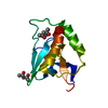
| ||||||||
|---|---|---|---|---|---|---|---|---|---|
| 1 |
| ||||||||
| Unit cell |
|
-Components
| #1: Protein | Mass: 14825.825 Da / Num. of mol.: 1 Source method: isolated from a genetically manipulated source Source: (gene. exp.) Homo sapiens (human) / Gene: PLAAT3, HRASLS3, HREV107, PLA2G16 / Production host:  References: UniProt: P53816, Transferases; Acyltransferases; Transferring groups other than aminoacyl groups, phospholipase A1, phospholipase A2 | ||||
|---|---|---|---|---|---|
| #2: Chemical |   Mass: 118.174 Da / Num. of mol.: 3 / Source method: obtained synthetically / Formula: C6H14O2 / Comment: precipitant*YM Mass: 118.174 Da / Num. of mol.: 3 / Source method: obtained synthetically / Formula: C6H14O2 / Comment: precipitant*YM#3: Water | ChemComp-HOH / |  Mass: 18.015 Da / Num. of mol.: 51 / Source method: isolated from a natural source / Formula: H2O Mass: 18.015 Da / Num. of mol.: 51 / Source method: isolated from a natural source / Formula: H2OHas ligand of interest | N | |
-Experimental details
-Experiment
| Experiment | Method: X-RAY DIFFRACTION / Number of used crystals: 1 |
|---|
- Sample preparation
| Crystal | Density Matthews: 2.65 Å3/Da / Density % sol: 53.61 % |
|---|---|
| Crystal grow | Temperature: 277 K / Method: vapor diffusion, sitting drop / Details: 0.1M Hepes-NaOH pH7.0-7.5, 45-70% MPD |
-Data collection
| Diffraction | Mean temperature: 100 K / Serial crystal experiment: N |
|---|---|
| Diffraction source | Source: SYNCHROTRON / Site: ESRF  / Beamline: ID23-1 / Wavelength: 0.97632 Å / Beamline: ID23-1 / Wavelength: 0.97632 Å |
| Detector | Type: DECTRIS PILATUS 6M / Detector: PIXEL / Date: Sep 27, 2010 |
| Radiation | Protocol: SINGLE WAVELENGTH / Monochromatic (M) / Laue (L): M / Scattering type: x-ray |
| Radiation wavelength | Wavelength: 0.97632 Å / Relative weight: 1 |
| Reflection | Resolution: 1.6→42.84 Å / Num. obs: 21212 / % possible obs: 99.7 % / Redundancy: 6.4 % / Rmerge(I) obs: 0.033 / Rpim(I) all: 0.021 / Rrim(I) all: 0.039 / Net I/σ(I): 21.4 |
| Reflection shell | Resolution: 1.6→1.63 Å / Rmerge(I) obs: 0.651 / Mean I/σ(I) obs: 2.5 / Num. unique obs: 999 / Rpim(I) all: 0.423 / Rrim(I) all: 0.779 / % possible all: 97.3 |
- Processing
| Software |
| |||||||||||||||||||||||||||||||||||||||||||||||||||||||||||||||||||||||||||||||||||||||||||||||||||||||||||||||||||||||||||||||||||||||||||||||||||
|---|---|---|---|---|---|---|---|---|---|---|---|---|---|---|---|---|---|---|---|---|---|---|---|---|---|---|---|---|---|---|---|---|---|---|---|---|---|---|---|---|---|---|---|---|---|---|---|---|---|---|---|---|---|---|---|---|---|---|---|---|---|---|---|---|---|---|---|---|---|---|---|---|---|---|---|---|---|---|---|---|---|---|---|---|---|---|---|---|---|---|---|---|---|---|---|---|---|---|---|---|---|---|---|---|---|---|---|---|---|---|---|---|---|---|---|---|---|---|---|---|---|---|---|---|---|---|---|---|---|---|---|---|---|---|---|---|---|---|---|---|---|---|---|---|---|---|---|---|
| Refinement | Method to determine structure: MIRAS / Resolution: 1.601→42.84 Å / Cor.coef. Fo:Fc: 0.967 / Cor.coef. Fo:Fc free: 0.958 / Cross valid method: FREE R-VALUE / ESU R: 0.074 / ESU R Free: 0.076 Details: Hydrogens have been used if present in the input file
| |||||||||||||||||||||||||||||||||||||||||||||||||||||||||||||||||||||||||||||||||||||||||||||||||||||||||||||||||||||||||||||||||||||||||||||||||||
| Solvent computation | Ion probe radii: 0.8 Å / Shrinkage radii: 0.8 Å / VDW probe radii: 1.2 Å / Solvent model: BABINET MODEL PLUS MASK | |||||||||||||||||||||||||||||||||||||||||||||||||||||||||||||||||||||||||||||||||||||||||||||||||||||||||||||||||||||||||||||||||||||||||||||||||||
| Displacement parameters | Biso mean: 38.16 Å2
| |||||||||||||||||||||||||||||||||||||||||||||||||||||||||||||||||||||||||||||||||||||||||||||||||||||||||||||||||||||||||||||||||||||||||||||||||||
| Refinement step | Cycle: LAST / Resolution: 1.601→42.84 Å
| |||||||||||||||||||||||||||||||||||||||||||||||||||||||||||||||||||||||||||||||||||||||||||||||||||||||||||||||||||||||||||||||||||||||||||||||||||
| Refine LS restraints |
| |||||||||||||||||||||||||||||||||||||||||||||||||||||||||||||||||||||||||||||||||||||||||||||||||||||||||||||||||||||||||||||||||||||||||||||||||||
| LS refinement shell |
|
