 Movie
Movie Controller

[English] 日本語
 Yorodumi
- PDB-7zoh: Carbohydrate binding domain CBM92-B from a multi-catalytic glucan... -
+ Open data

- Basic information
| Entry | Database: PDB / ID: 7zoh | |||||||||
|---|---|---|---|---|---|---|---|---|---|---|
| Title | Carbohydrate binding domain CBM92-B from a multi-catalytic glucanase-chitinase from Chitinophaga pinensis DSM 2588 | |||||||||
 Components Components | Glycoside hydrolase family 18 | |||||||||
 Keywords Keywords | CARBOHYDRATE / beta-trefoil / carbohydrate binding domain / beta-glycan | |||||||||
| Function / homology |  Function and homology information Function and homology informationglucan catabolic process / beta-glucosidase activity / chitin binding / cell surface / extracellular region Similarity search - Function | |||||||||
| Biological species |  Chitinophaga pinensis DSM 2588 (bacteria) Chitinophaga pinensis DSM 2588 (bacteria) | |||||||||
| Method |  X-RAY DIFFRACTION / SYNCHROTRON / MOLECULAR REPLACEMENT / Resolution: 1.56 Å X-RAY DIFFRACTION / SYNCHROTRON / MOLECULAR REPLACEMENT / Resolution: 1.56 Å | |||||||||
 Authors Authors | Mazurkewich, S. / McKee, L.S. / Lu, Z. / Branden, G. / Larsbrink, J. | |||||||||
| Funding support |  Sweden, 2items Sweden, 2items
| |||||||||
 Citation Citation | Journal: Nat Commun / Year: 2024 Title: Structural and biochemical analysis of family 92 carbohydrate-binding modules uncovers multivalent binding to beta-glucans. Authors: Hao, M.S. / Mazurkewich, S. / Li, H. / Kvammen, A. / Saha, S. / Koskela, S. / Inman, A.R. / Nakajima, M. / Tanaka, N. / Nakai, H. / Branden, G. / Bulone, V. / Larsbrink, J. / McKee, L.S. | |||||||||
| History |
|
- Structure visualization
| Structure viewer | Molecule: MolmilJmol/JSmol |
|---|
- Downloads & links
-Download
| PDBx/mmCIF format | 7zoh.cif.gz | 122.9 KB | Display | PDBx/mmCIF format |
|---|---|---|---|---|
| PDB format | pdb7zoh.ent.gz | 91.3 KB | Display | PDB format |
| PDBx/mmJSON format | 7zoh.json.gz | Tree view | PDBx/mmJSON format | |
| Others |  Other downloads Other downloads |
-Validation report
| Arichive directory | https://data.pdbj.org/pub/pdb/validation_reports/zo/7zohftp://data.pdbj.org/pub/pdb/validation_reports/zo/7zoh | HTTPS FTP |
|---|
-Related structure data
| Related structure data |  7zoiC  7zonC 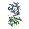 7zooC 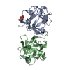 7zopC  3llpS C: citing same article ( S: Starting model for refinement |
|---|---|
| Similar structure data |
-Links
 PDBj
PDBj- Assembly
| Deposited unit | 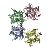
| ||||||||||||
|---|---|---|---|---|---|---|---|---|---|---|---|---|---|
| 1 | 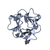
| ||||||||||||
| 2 | 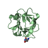
| ||||||||||||
| 3 | 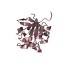
| ||||||||||||
| 4 | 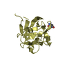
| ||||||||||||
| Unit cell |
|
-Components
| #1: Protein | Mass: 16176.707 Da / Num. of mol.: 4 Source method: isolated from a genetically manipulated source Details: succinimide observed substituted at Asp Source: (gene. exp.) Chitinophaga pinensis DSM 2588 (bacteria)Gene: Cpin_2580 / Production host: #2: Water | ChemComp-HOH / |  Mass: 18.015 Da / Num. of mol.: 313 / Source method: isolated from a natural source / Formula: H2O Mass: 18.015 Da / Num. of mol.: 313 / Source method: isolated from a natural source / Formula: H2OHas ligand of interest | N | Has protein modification | Y | |
|---|
-Experimental details
-Experiment
| Experiment | Method: X-RAY DIFFRACTION / Number of used crystals: 1 |
|---|
- Sample preparation
| Crystal | Density Matthews: 2.03 Å3/Da / Density % sol: 39.49 % |
|---|---|
| Crystal grow | Temperature: 293 K / Method: vapor diffusion, sitting drop / Details: 0.1 M citrate buffer pH 5.5 with 15 % w/v PEG 3350 |
-Data collection
| Diffraction | Mean temperature: 100 K / Serial crystal experiment: N |
|---|---|
| Diffraction source | Source: SYNCHROTRON / Site: MAX IV / Beamline: BioMAX / Wavelength: 1.0332 Å |
| Detector | Type: DECTRIS EIGER X 16M / Detector: PIXEL / Date: Mar 27, 2020 |
| Radiation | Protocol: SINGLE WAVELENGTH / Monochromatic (M) / Laue (L): M / Scattering type: x-ray |
| Radiation wavelength | Wavelength: 1.0332 Å / Relative weight: 1 |
| Reflection | Resolution: 1.56→33.97 Å / Num. obs: 69506 / % possible obs: 95.35 % / Redundancy: 3.6 % / Biso Wilson estimate: 24.42 Å2 / CC1/2: 0.996 / Rmerge(I) obs: 0.075 / Net I/σ(I): 8.74 |
| Reflection shell | Resolution: 1.56→1.61 Å / Redundancy: 3.4 % / Rmerge(I) obs: 1.13 / Mean I/σ(I) obs: 1.04 / Num. unique obs: 6543 / CC1/2: 0.415 / % possible all: 88.81 |
- Processing
| Software |
| |||||||||||||||||||||||||||||||||||||||||||||||||||||||||||||||||||||||||||||||||||||||||||||||||||||||||||||||||||||||||||||||||||||||||||||||||||||||||||||||||||||||||||||||||||||||||||||||||||||||||||||||||||||||||
|---|---|---|---|---|---|---|---|---|---|---|---|---|---|---|---|---|---|---|---|---|---|---|---|---|---|---|---|---|---|---|---|---|---|---|---|---|---|---|---|---|---|---|---|---|---|---|---|---|---|---|---|---|---|---|---|---|---|---|---|---|---|---|---|---|---|---|---|---|---|---|---|---|---|---|---|---|---|---|---|---|---|---|---|---|---|---|---|---|---|---|---|---|---|---|---|---|---|---|---|---|---|---|---|---|---|---|---|---|---|---|---|---|---|---|---|---|---|---|---|---|---|---|---|---|---|---|---|---|---|---|---|---|---|---|---|---|---|---|---|---|---|---|---|---|---|---|---|---|---|---|---|---|---|---|---|---|---|---|---|---|---|---|---|---|---|---|---|---|---|---|---|---|---|---|---|---|---|---|---|---|---|---|---|---|---|---|---|---|---|---|---|---|---|---|---|---|---|---|---|---|---|---|---|---|---|---|---|---|---|---|---|---|---|---|---|---|---|---|
| Refinement | Method to determine structure: MOLECULAR REPLACEMENT Starting model: 3LLP Resolution: 1.56→33.97 Å / SU ML: 0.1833 / Cross valid method: FREE R-VALUE / σ(F): 1.96 / Phase error: 21.826 Stereochemistry target values: GeoStd + Monomer Library + CDL v1.2
| |||||||||||||||||||||||||||||||||||||||||||||||||||||||||||||||||||||||||||||||||||||||||||||||||||||||||||||||||||||||||||||||||||||||||||||||||||||||||||||||||||||||||||||||||||||||||||||||||||||||||||||||||||||||||
| Solvent computation | Shrinkage radii: 0.9 Å / VDW probe radii: 1.11 Å / Solvent model: FLAT BULK SOLVENT MODEL | |||||||||||||||||||||||||||||||||||||||||||||||||||||||||||||||||||||||||||||||||||||||||||||||||||||||||||||||||||||||||||||||||||||||||||||||||||||||||||||||||||||||||||||||||||||||||||||||||||||||||||||||||||||||||
| Displacement parameters | Biso mean: 30.11 Å2 | |||||||||||||||||||||||||||||||||||||||||||||||||||||||||||||||||||||||||||||||||||||||||||||||||||||||||||||||||||||||||||||||||||||||||||||||||||||||||||||||||||||||||||||||||||||||||||||||||||||||||||||||||||||||||
| Refinement step | Cycle: LAST / Resolution: 1.56→33.97 Å
| |||||||||||||||||||||||||||||||||||||||||||||||||||||||||||||||||||||||||||||||||||||||||||||||||||||||||||||||||||||||||||||||||||||||||||||||||||||||||||||||||||||||||||||||||||||||||||||||||||||||||||||||||||||||||
| Refine LS restraints |
| |||||||||||||||||||||||||||||||||||||||||||||||||||||||||||||||||||||||||||||||||||||||||||||||||||||||||||||||||||||||||||||||||||||||||||||||||||||||||||||||||||||||||||||||||||||||||||||||||||||||||||||||||||||||||
| LS refinement shell |
|
