 Movie
Movie Controller


+ Open data

- Basic information
| Entry | Database: PDB / ID: 7zn1 | ||||||
|---|---|---|---|---|---|---|---|
| Title | Avidin + Biotin-Tempo | ||||||
 Components Components | Avidin | ||||||
 Keywords Keywords | PROTEIN BINDING / Avidin / Biotin / TEMPO free radical | ||||||
| Function / homology |  Function and homology information Function and homology information | ||||||
| Biological species |  | ||||||
| Method |  X-RAY DIFFRACTION / SYNCHROTRON / MOLECULAR REPLACEMENT / Resolution: 2.33 Å X-RAY DIFFRACTION / SYNCHROTRON / MOLECULAR REPLACEMENT / Resolution: 2.33 Å | ||||||
 Authors Authors | Milani, J. / Lau, K. / Pojer, F. / Ansermet, J.P. / Saenz, F. | ||||||
| Funding support | 1items
| ||||||
 Citation Citation | Journal: To Be Published Title: Avidin + Biotin-Tempo Authors: Milani, J. / Lau, K. / Pojer, F. / Ansermet, J.P. / Saenz, F. | ||||||
| History |
|
- Structure visualization
| Structure viewer | Molecule: MolmilJmol/JSmol |
|---|
- Downloads & links
-Download
| PDBx/mmCIF format | 7zn1.cif.gz | 141.2 KB | Display | PDBx/mmCIF format |
|---|---|---|---|---|
| PDB format | pdb7zn1.ent.gz | 89.4 KB | Display | PDB format |
| PDBx/mmJSON format | 7zn1.json.gz | Tree view | PDBx/mmJSON format | |
| Others |  Other downloads Other downloads |
-Validation report
| Arichive directory | https://data.pdbj.org/pub/pdb/validation_reports/zn/7zn1ftp://data.pdbj.org/pub/pdb/validation_reports/zn/7zn1 | HTTPS FTP |
|---|
-Related structure data
| Related structure data | 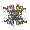 7zylC  1ravS C: citing same article ( S: Starting model for refinement |
|---|---|
| Similar structure data |
-Links
 PDBj
PDBj- Assembly
| Deposited unit | 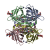
| ||||||||||||
|---|---|---|---|---|---|---|---|---|---|---|---|---|---|
| 1 |
| ||||||||||||
| Unit cell |
|
-Components
-Protein , 1 types, 4 molecules ABCD
| #1: Protein | Mass: 16787.193 Da / Num. of mol.: 4 / Source method: isolated from a natural source / Source: (natural) |
|---|
-Sugars , 2 types, 4 molecules 
| #2: Polysaccharide | 2-acetamido-2-deoxy-beta-D-glucopyranose-(1-4)-2-acetamido-2-deoxy-beta-D-glucopyranose Source method: isolated from a genetically manipulated source |
|---|---|
| #3: Sugar |  Type: D-saccharide, beta linking / Mass: 221.208 Da / Num. of mol.: 3 / Source method: obtained synthetically / Formula: C8H15NO6 Type: D-saccharide, beta linking / Mass: 221.208 Da / Num. of mol.: 3 / Source method: obtained synthetically / Formula: C8H15NO6 |
-Non-polymers , 4 types, 55 molecules 


| #4: Chemical | ChemComp-KFL /  Mass: 412.590 Da / Num. of mol.: 4 / Source method: obtained synthetically / Formula: C20H36N4O3S / Feature type: SUBJECT OF INVESTIGATION Mass: 412.590 Da / Num. of mol.: 4 / Source method: obtained synthetically / Formula: C20H36N4O3S / Feature type: SUBJECT OF INVESTIGATION#5: Chemical | ChemComp-ZN /  Mass: 65.409 Da / Num. of mol.: 4 / Source method: obtained synthetically / Formula: Zn Mass: 65.409 Da / Num. of mol.: 4 / Source method: obtained synthetically / Formula: Zn#6: Chemical | ChemComp-NA /  Mass: 22.990 Da / Num. of mol.: 4 / Source method: isolated from a natural source / Formula: Na Mass: 22.990 Da / Num. of mol.: 4 / Source method: isolated from a natural source / Formula: Na#7: Water | ChemComp-HOH / | Mass: 18.015 Da / Num. of mol.: 43 / Source method: isolated from a natural source / Formula: H2O |
|---|
-Details
| Has ligand of interest | Y |
|---|---|
| Has protein modification | Y |
-Experimental details
-Experiment
| Experiment | Method: X-RAY DIFFRACTION / Number of used crystals: 1 |
|---|
- Sample preparation
| Crystal | Density Matthews: 1.91 Å3/Da / Density % sol: 35.46 % |
|---|---|
| Crystal grow | Temperature: 298.15 K / Method: vapor diffusion, sitting drop / Details: 0.2 M Zinc Acetate dihydrate 22% w/v PEG3350 |
-Data collection
| Diffraction | Mean temperature: 100 K / Serial crystal experiment: N |
|---|---|
| Diffraction source | Source: SYNCHROTRON / Site: SLS  / Beamline: X06DA / Wavelength: 1 Å / Beamline: X06DA / Wavelength: 1 Å |
| Detector | Type: DECTRIS PILATUS 2M-F / Detector: PIXEL / Date: Dec 10, 2021 |
| Radiation | Protocol: SINGLE WAVELENGTH / Monochromatic (M) / Laue (L): M / Scattering type: x-ray |
| Radiation wavelength | Wavelength: 1 Å / Relative weight: 1 |
| Reflection | Resolution: 2.33→44.5 Å / Num. obs: 40645 / % possible obs: 95.9 % / Redundancy: 2.13 % / Biso Wilson estimate: 44.79 Å2 / CC1/2: 0.995 / Net I/σ(I): 5.13 |
| Reflection shell | Resolution: 2.33→2.48 Å / Num. unique obs: 6570 / CC1/2: 0.609 |
- Processing
| Software |
| |||||||||||||||||||||||||||||||||||||||||||||||||||||||||||||||
|---|---|---|---|---|---|---|---|---|---|---|---|---|---|---|---|---|---|---|---|---|---|---|---|---|---|---|---|---|---|---|---|---|---|---|---|---|---|---|---|---|---|---|---|---|---|---|---|---|---|---|---|---|---|---|---|---|---|---|---|---|---|---|---|---|
| Refinement | Method to determine structure: MOLECULAR REPLACEMENT Starting model: 1rav Resolution: 2.33→44.5 Å / SU ML: 0.3528 / Cross valid method: FREE R-VALUE / σ(F): 1.35 / Phase error: 36.7179 Stereochemistry target values: GeoStd + Monomer Library + CDL v1.2
| |||||||||||||||||||||||||||||||||||||||||||||||||||||||||||||||
| Solvent computation | Shrinkage radii: 0.9 Å / VDW probe radii: 1.1 Å / Solvent model: FLAT BULK SOLVENT MODEL | |||||||||||||||||||||||||||||||||||||||||||||||||||||||||||||||
| Displacement parameters | Biso mean: 50.84 Å2 | |||||||||||||||||||||||||||||||||||||||||||||||||||||||||||||||
| Refinement step | Cycle: LAST / Resolution: 2.33→44.5 Å
| |||||||||||||||||||||||||||||||||||||||||||||||||||||||||||||||
| Refine LS restraints |
| |||||||||||||||||||||||||||||||||||||||||||||||||||||||||||||||
| LS refinement shell |
|
