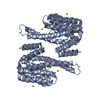
- PDB-7zmu: 14-3-3s binding to non-natural peptide 2d -
+
データを開く
IDまたはキーワード:
読み込み中...
-
基本情報
登録情報
データベース: PDB / ID: 7zmu
タイトル
14-3-3s binding to non-natural peptide 2d
要素
14-3-3 protein sigma
non-natural peptide 2
キーワード
PEPTIDE BINDING PROTEIN / 14-3-3 / non-natural peptide
機能・相同性
機能・相同性情報
regulation of epidermal cell division / protein kinase C inhibitor activity / positive regulation of epidermal cell differentiation / keratinocyte development / keratinization / regulation of cell-cell adhesion / cAMP/PKA signal transduction / Regulation of localization of FOXO transcription factors / keratinocyte proliferation / phosphoserine residue binding ...regulation of epidermal cell division / protein kinase C inhibitor activity / positive regulation of epidermal cell differentiation / keratinocyte development / keratinization / regulation of cell-cell adhesion / cAMP/PKA signal transduction / Regulation of localization of FOXO transcription factors / keratinocyte proliferation / phosphoserine residue binding / Activation of BAD and translocation to mitochondria / negative regulation of keratinocyte proliferation / establishment of skin barrier / negative regulation of protein localization to plasma membrane / Chk1/Chk2(Cds1) mediated inactivation of Cyclin B:Cdk1 complex / SARS-CoV-2 targets host intracellular signalling and regulatory pathways / negative regulation of stem cell proliferation / SARS-CoV-1 targets host intracellular signalling and regulatory pathways / RHO GTPases activate PKNs / positive regulation of protein localization / positive regulation of cell adhesion / protein sequestering activity / negative regulation of innate immune response / protein export from nucleus / release of cytochrome c from mitochondria / TP53 Regulates Transcription of Genes Involved in G2 Cell Cycle Arrest / positive regulation of protein export from nucleus / negative regulation of protein kinase activity / stem cell proliferation / Translocation of SLC2A4 (GLUT4) to the plasma membrane / TP53 Regulates Metabolic Genes / intrinsic apoptotic signaling pathway in response to DNA damage / intracellular protein localization / regulation of protein localization / positive regulation of cell growth / regulation of cell cycle / cadherin binding / protein kinase binding / negative regulation of transcription by RNA polymerase II / signal transduction / extracellular space / extracellular exosome / identical protein binding / nucleus / cytosol / cytoplasm 類似検索 - 分子機能
14-3-3proteinsigma / Epithelial cell marker protein 1 / Stratifin
分子量: 26542.914 Da / 分子数: 1 / 由来タイプ: 組換発現 詳細: The initial GAMGS amino acids form the expression tag of the protein 由来: (組換発現) Homo sapiens (ヒト) / 遺伝子: SFN, HME1 / 発現宿主: Escherichia coli (大腸菌) / 参照: UniProt: P31947
 ムービー
ムービー コントローラー


 データを開く
 基本情報
基本情報 要素
要素 キーワード
キーワード 機能・相同性情報
 Homo sapiens (ヒト)
 X線回折 /
  データ登録者
 オランダ, 1件
  引用
引用 構造の表示
構造の表示 ダウンロードとリンク
 その他のダウンロード
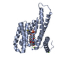
 PDBj
PDBj


 集合体
集合体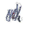


 分子量: 24.305 Da / 分子数: 2 / 由来タイプ: 合成 / 式: Mg
 分子量: 18.015 Da / 分子数: 253 / 由来タイプ: 天然 / 式: H2O
 試料調製
試料調製 / ビームライン: P11 / 波長: 1.0332 Å
 解析
解析