 Movie
Movie Controller


+ Open data

- Basic information
| Entry | Database: PDB / ID: 7zmw | |||||||||
|---|---|---|---|---|---|---|---|---|---|---|
| Title | 14-3-3s binding to non-natural peptide 2c | |||||||||
 Components Components |
| |||||||||
 Keywords Keywords | PEPTIDE BINDING PROTEIN / 14-3-3 / non-natural peptide | |||||||||
| Function / homology |  Function and homology information Function and homology informationpositive regulation of epidermal cell differentiation / keratinocyte development / regulation of epidermal cell division / protein kinase C inhibitor activity / keratinization / regulation of cell-cell adhesion / keratinocyte proliferation / establishment of skin barrier / Regulation of localization of FOXO transcription factors / negative regulation of keratinocyte proliferation ...positive regulation of epidermal cell differentiation / keratinocyte development / regulation of epidermal cell division / protein kinase C inhibitor activity / keratinization / regulation of cell-cell adhesion / keratinocyte proliferation / establishment of skin barrier / Regulation of localization of FOXO transcription factors / negative regulation of keratinocyte proliferation / phosphoserine residue binding / Activation of BAD and translocation to mitochondria / cAMP/PKA signal transduction / negative regulation of stem cell proliferation / negative regulation of protein localization to plasma membrane / SARS-CoV-2 targets host intracellular signalling and regulatory pathways / negative regulation of protein kinase activity / Chk1/Chk2(Cds1) mediated inactivation of Cyclin B:Cdk1 complex / SARS-CoV-1 targets host intracellular signalling and regulatory pathways / RHO GTPases activate PKNs / positive regulation of protein localization / stem cell proliferation / protein export from nucleus / release of cytochrome c from mitochondria / TP53 Regulates Transcription of Genes Involved in G2 Cell Cycle Arrest / negative regulation of innate immune response / positive regulation of cell adhesion / positive regulation of protein export from nucleus / TP53 Regulates Metabolic Genes / Translocation of SLC2A4 (GLUT4) to the plasma membrane / protein sequestering activity / intrinsic apoptotic signaling pathway in response to DNA damage / intracellular protein localization / regulation of protein localization / sperm midpiece / positive regulation of cell growth / regulation of cell cycle / cadherin binding / protein kinase binding / negative regulation of transcription by RNA polymerase II / signal transduction / : / extracellular exosome / identical protein binding / nucleus / cytosol / cytoplasm Similarity search - Function | |||||||||
| Biological species |  Homo sapiens (human) Homo sapiens (human)synthetic construct (others) | |||||||||
| Method |  X-RAY DIFFRACTION / MOLECULAR REPLACEMENT / Resolution: 1.8 Å X-RAY DIFFRACTION / MOLECULAR REPLACEMENT / Resolution: 1.8 Å | |||||||||
 Authors Authors | Somsen, B.A. / Craenmehr, F.W.B. / Ottmann, C. | |||||||||
| Funding support |  Netherlands, 1items Netherlands, 1items
| |||||||||
 Citation Citation | Journal: Chem Sci / Year: 2022 Title: Functional mapping of the 14-3-3 hub protein as a guide to design 14-3-3 molecular glues. Authors: Somsen, B.A. / Craenmehr, F.W.B. / Liu, W.W. / Koops, A.A. / Pennings, M.A.M. / Visser, E.J. / Ottmann, C. / Cossar, P.J. / Brunsveld, L. | |||||||||
| History |
|
- Structure visualization
| Structure viewer | Molecule: MolmilJmol/JSmol |
|---|
- Downloads & links
-Download
| PDBx/mmCIF format | 7zmw.cif.gz | 111.4 KB | Display | PDBx/mmCIF format |
|---|---|---|---|---|
| PDB format | pdb7zmw.ent.gz | 84.8 KB | Display | PDB format |
| PDBx/mmJSON format | 7zmw.json.gz | Tree view | PDBx/mmJSON format | |
| Others |  Other downloads Other downloads |
-Validation report
| Arichive directory | https://data.pdbj.org/pub/pdb/validation_reports/zm/7zmwftp://data.pdbj.org/pub/pdb/validation_reports/zm/7zmw | HTTPS FTP |
|---|
-Related structure data
| Related structure data | 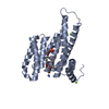 7zmuC 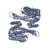 4jc3S S: Starting model for refinement C: citing same article ( |
|---|---|
| Similar structure data |
-Links
 PDBj
PDBj


- Assembly
| Deposited unit | 
| ||||||||
|---|---|---|---|---|---|---|---|---|---|
| 1 | 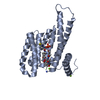
| ||||||||
| Unit cell |
| ||||||||
| Components on special symmetry positions |
|
-Components
| #1: Protein | Mass: 26542.914 Da / Num. of mol.: 1 Source method: isolated from a genetically manipulated source Details: The initial GAMGS amino acids form the expression tag of the protein Source: (gene. exp.) Homo sapiens (human) / Gene: SFN, HME1 / Production host:  | ||||
|---|---|---|---|---|---|
| #2: Protein/peptide | Mass: 1022.975 Da / Num. of mol.: 1 / Source method: obtained synthetically / Source: (synth.) synthetic construct (others) | ||||
| #3: Chemical |   Mass: 24.305 Da / Num. of mol.: 2 / Source method: obtained synthetically / Formula: Mg Mass: 24.305 Da / Num. of mol.: 2 / Source method: obtained synthetically / Formula: Mg#4: Water | ChemComp-HOH / |  Mass: 18.015 Da / Num. of mol.: 265 / Source method: isolated from a natural source / Formula: H2O Mass: 18.015 Da / Num. of mol.: 265 / Source method: isolated from a natural source / Formula: H2OHas ligand of interest | Y | |
-Experimental details
-Experiment
| Experiment | Method: X-RAY DIFFRACTION / Number of used crystals: 1 |
|---|
- Sample preparation
| Crystal | Density Matthews: 2.69 Å3/Da / Density % sol: 54.31 % |
|---|---|
| Crystal grow | Temperature: 277 K / Method: vapor diffusion, hanging drop / pH: 7.5 Details: 95 mM HEPES pH 7.5, 0.19 M CaCl2, 5% glycerol and 28% PEG400 |
-Data collection
| Diffraction | Mean temperature: 100 K / Serial crystal experiment: N |
|---|---|
| Diffraction source | Source: SEALED TUBE / Type: RIGAKU MICROMAX-003 / Wavelength: 1.54187 Å |
| Detector | Type: DECTRIS PILATUS 200K / Detector: PIXEL / Date: Jan 20, 2021 |
| Radiation | Protocol: SINGLE WAVELENGTH / Monochromatic (M) / Laue (L): M / Scattering type: x-ray |
| Radiation wavelength | Wavelength: 1.54187 Å / Relative weight: 1 |
| Reflection | Resolution: 1.8→33.03 Å / Num. obs: 48998 / % possible obs: 97.4 % / Redundancy: 6 % / CC1/2: 0.996 / Net I/σ(I): 11.2 |
| Reflection shell | Resolution: 1.8→1.84 Å / Mean I/σ(I) obs: 3 / Num. unique obs: 1415 / CC1/2: 0.893 |
- Processing
| Software |
| |||||||||||||||||||||||||||||||||||||||||||||||||||||||||||||||||||||||||||||||||||||||||||||||||||||||||||||||||||||||||||||||||||||
|---|---|---|---|---|---|---|---|---|---|---|---|---|---|---|---|---|---|---|---|---|---|---|---|---|---|---|---|---|---|---|---|---|---|---|---|---|---|---|---|---|---|---|---|---|---|---|---|---|---|---|---|---|---|---|---|---|---|---|---|---|---|---|---|---|---|---|---|---|---|---|---|---|---|---|---|---|---|---|---|---|---|---|---|---|---|---|---|---|---|---|---|---|---|---|---|---|---|---|---|---|---|---|---|---|---|---|---|---|---|---|---|---|---|---|---|---|---|---|---|---|---|---|---|---|---|---|---|---|---|---|---|---|---|---|
| Refinement | Method to determine structure: MOLECULAR REPLACEMENT Starting model: 4jc3 Resolution: 1.8→33.03 Å / SU ML: 0.19 / Cross valid method: THROUGHOUT / σ(F): 1.34 / Phase error: 21.55 / Stereochemistry target values: ML
| |||||||||||||||||||||||||||||||||||||||||||||||||||||||||||||||||||||||||||||||||||||||||||||||||||||||||||||||||||||||||||||||||||||
| Solvent computation | Shrinkage radii: 0.9 Å / VDW probe radii: 1.1 Å / Solvent model: FLAT BULK SOLVENT MODEL | |||||||||||||||||||||||||||||||||||||||||||||||||||||||||||||||||||||||||||||||||||||||||||||||||||||||||||||||||||||||||||||||||||||
| Displacement parameters | Biso max: 78.51 Å2 / Biso mean: 14.4028 Å2 / Biso min: 0.08 Å2 | |||||||||||||||||||||||||||||||||||||||||||||||||||||||||||||||||||||||||||||||||||||||||||||||||||||||||||||||||||||||||||||||||||||
| Refinement step | Cycle: final / Resolution: 1.8→33.03 Å
| |||||||||||||||||||||||||||||||||||||||||||||||||||||||||||||||||||||||||||||||||||||||||||||||||||||||||||||||||||||||||||||||||||||
| LS refinement shell | Refine-ID: X-RAY DIFFRACTION / Rfactor Rfree error: 0 / Total num. of bins used: 18
|
