 Movie
Movie Controller


+ Open data

- Basic information
| Entry | Database: PDB / ID: 7zkx | ||||||
|---|---|---|---|---|---|---|---|
| Title | SRPK2 IN COMPLEX WITH INHIBITOR | ||||||
 Components Components | SRSF protein kinase 2 | ||||||
 Keywords Keywords | TRANSFERASE / SERINE/THREONINE-PROTEIN KINASE / SRPK2 / NUCLEAR PROTEIN | ||||||
| Function / homology |  Function and homology information Function and homology informationnuclear speck organization / R-loop processing / regulation of mRNA processing / regulation of mRNA splicing, via spliceosome / Maturation of nucleoprotein / negative regulation of viral genome replication / spliceosomal complex assembly / positive regulation of viral genome replication / sperm end piece / 14-3-3 protein binding ...nuclear speck organization / R-loop processing / regulation of mRNA processing / regulation of mRNA splicing, via spliceosome / Maturation of nucleoprotein / negative regulation of viral genome replication / spliceosomal complex assembly / positive regulation of viral genome replication / sperm end piece / 14-3-3 protein binding / positive regulation of cell cycle / acrosomal vesicle / RNA splicing / phosphatidylinositol 3-kinase/protein kinase B signal transduction / peptidyl-serine phosphorylation / positive regulation of neuron apoptotic process / presynapse / angiogenesis / cell differentiation / protein phosphorylation / non-specific serine/threonine protein kinase / intracellular signal transduction / nuclear speck / innate immune response / protein serine kinase activity / protein serine/threonine kinase activity / positive regulation of cell population proliferation / positive regulation of gene expression / chromatin / nucleolus / magnesium ion binding / RNA binding / nucleoplasm / ATP binding / nucleus / cytoplasm / cytosol Similarity search - Function | ||||||
| Biological species |  Homo sapiens (human) Homo sapiens (human) | ||||||
| Method |  X-RAY DIFFRACTION / SYNCHROTRON / MOLECULAR REPLACEMENT / molecular replacement / Resolution: 2.06 Å X-RAY DIFFRACTION / SYNCHROTRON / MOLECULAR REPLACEMENT / molecular replacement / Resolution: 2.06 Å | ||||||
 Authors Authors | Graedler, U. | ||||||
| Funding support | 1items
| ||||||
 Citation Citation | Journal: J.Med.Chem. / Year: 2023 Title: MSC-1186, a Highly Selective Pan-SRPK Inhibitor Based on an Exceptionally Decorated Benzimidazole-Pyrimidine Core. Authors: Schroder, M. / Leiendecker, M. / Gradler, U. / Braun, J. / Blum, A. / Wanior, M. / Berger, B.T. / Kramer, A. / Muller, S. / Esdar, C. / Knapp, S. / Heinrich, T. | ||||||
| History |
|
- Structure visualization
| Structure viewer | Molecule: MolmilJmol/JSmol |
|---|
- Downloads & links
-Download
| PDBx/mmCIF format | 7zkx.cif.gz | 96.9 KB | Display | PDBx/mmCIF format |
|---|---|---|---|---|
| PDB format | pdb7zkx.ent.gz | 68 KB | Display | PDB format |
| PDBx/mmJSON format | 7zkx.json.gz | Tree view | PDBx/mmJSON format | |
| Others |  Other downloads Other downloads |
-Validation report
| Arichive directory | https://data.pdbj.org/pub/pdb/validation_reports/zk/7zkxftp://data.pdbj.org/pub/pdb/validation_reports/zk/7zkx | HTTPS FTP |
|---|
-Related structure data
| Related structure data |  7zksC 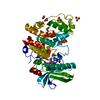 2x7gS C: citing same article ( S: Starting model for refinement |
|---|---|
| Similar structure data |
-Links
 PDBj
PDBj- Assembly
| Deposited unit | 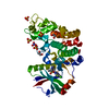
| ||||||||
|---|---|---|---|---|---|---|---|---|---|
| 1 |
| ||||||||
| Unit cell |
|
-Components
-Protein , 1 types, 1 molecules A
| #1: Protein | Mass: 70206.562 Da / Num. of mol.: 1 / Fragment: KINASE DOMAIN Source method: isolated from a genetically manipulated source Source: (gene. exp.) Homo sapiens (human) / Gene: SRPK2 / Production host:  References: UniProt: P78362, non-specific serine/threonine protein kinase |
|---|
-Non-polymers , 6 types, 145 molecules 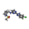


| #2: Chemical | ChemComp-IXQ /  Mass: 461.900 Da / Num. of mol.: 1 / Source method: obtained synthetically / Formula: C19H17ClFN7O2S / Feature type: SUBJECT OF INVESTIGATION Mass: 461.900 Da / Num. of mol.: 1 / Source method: obtained synthetically / Formula: C19H17ClFN7O2S / Feature type: SUBJECT OF INVESTIGATION | ||||||||
|---|---|---|---|---|---|---|---|---|---|
| #3: Chemical | ChemComp-SO4 /  Mass: 96.063 Da / Num. of mol.: 5 / Source method: obtained synthetically / Formula: SO4 Mass: 96.063 Da / Num. of mol.: 5 / Source method: obtained synthetically / Formula: SO4#4: Chemical |  Mass: 62.068 Da / Num. of mol.: 3 / Source method: obtained synthetically / Formula: C2H6O2 Mass: 62.068 Da / Num. of mol.: 3 / Source method: obtained synthetically / Formula: C2H6O2#5: Chemical |  Mass: 62.005 Da / Num. of mol.: 2 / Source method: obtained synthetically / Formula: NO3 Mass: 62.005 Da / Num. of mol.: 2 / Source method: obtained synthetically / Formula: NO3#6: Chemical | ChemComp-ACT / |  Mass: 59.044 Da / Num. of mol.: 1 / Source method: obtained synthetically / Formula: C2H3O2 Mass: 59.044 Da / Num. of mol.: 1 / Source method: obtained synthetically / Formula: C2H3O2#7: Water | ChemComp-HOH / | Mass: 18.015 Da / Num. of mol.: 133 / Source method: isolated from a natural source / Formula: H2O |
-Details
| Has ligand of interest | Y |
|---|
-Experimental details
-Experiment
| Experiment | Method: X-RAY DIFFRACTION / Number of used crystals: 1 |
|---|
- Sample preparation
| Crystal | Density Matthews: 3.66 Å3/Da / Density % sol: 66.44 % |
|---|---|
| Crystal grow | Temperature: 293 K / Method: vapor diffusion / Details: 5 mM HEPES pH 7.5, 150 mM NaCl, 20 mM DTT |
-Data collection
| Diffraction | Mean temperature: 100 K / Serial crystal experiment: N |
|---|---|
| Diffraction source | Source: SYNCHROTRON / Site: SLS  / Beamline: X10SA / Wavelength: 0.99989 Å / Beamline: X10SA / Wavelength: 0.99989 Å |
| Detector | Type: DECTRIS PILATUS 6M / Detector: PIXEL / Date: Jan 16, 2019 |
| Radiation | Protocol: SINGLE WAVELENGTH / Monochromatic (M) / Laue (L): M / Scattering type: x-ray |
| Radiation wavelength | Wavelength: 0.99989 Å / Relative weight: 1 |
| Reflection | Resolution: 2.06→93.32 Å / Num. obs: 36972 / % possible obs: 97.9 % / Redundancy: 2.9 % / Rrim(I) all: 0.05 / Rsym value: 0.041 / Net I/σ(I): 15.51 |
| Reflection shell | Resolution: 2.06→2.31 Å / Redundancy: 2.9 % / Mean I/σ(I) obs: 2.53 / Num. unique obs: 10714 / Rrim(I) all: 0.584 / Rsym value: 0.473 / % possible all: 98.8 |
-Phasing
| Phasing | Method: molecular replacement |
|---|
- Processing
| Software |
| ||||||||||||||||||||||||||||||||||||||||||||||||||||||||||||
|---|---|---|---|---|---|---|---|---|---|---|---|---|---|---|---|---|---|---|---|---|---|---|---|---|---|---|---|---|---|---|---|---|---|---|---|---|---|---|---|---|---|---|---|---|---|---|---|---|---|---|---|---|---|---|---|---|---|---|---|---|---|
| Refinement | Method to determine structure: MOLECULAR REPLACEMENT Starting model: 2X7G Resolution: 2.06→93.32 Å / Cor.coef. Fo:Fc: 0.965 / Cor.coef. Fo:Fc free: 0.956 / SU B: 7.792 / SU ML: 0.102 / Cross valid method: THROUGHOUT / σ(F): 0 / ESU R: 0.134 / ESU R Free: 0.127 / Stereochemistry target values: MAXIMUM LIKELIHOOD
| ||||||||||||||||||||||||||||||||||||||||||||||||||||||||||||
| Solvent computation | Ion probe radii: 0.8 Å / Shrinkage radii: 0.8 Å / VDW probe radii: 1.2 Å / Solvent model: MASK | ||||||||||||||||||||||||||||||||||||||||||||||||||||||||||||
| Displacement parameters | Biso max: 129.33 Å2 / Biso mean: 57.306 Å2 / Biso min: 27.16 Å2
| ||||||||||||||||||||||||||||||||||||||||||||||||||||||||||||
| Refinement step | Cycle: final / Resolution: 2.06→93.32 Å
| ||||||||||||||||||||||||||||||||||||||||||||||||||||||||||||
| Refine LS restraints |
| ||||||||||||||||||||||||||||||||||||||||||||||||||||||||||||
| LS refinement shell | Resolution: 2.06→2.114 Å / Rfactor Rfree error: 0 / Total num. of bins used: 20
|
