 Movie
Movie Controller

[English] 日本語
 Yorodumi
- PDB-7zkh: C-Methyltransferase PsmD from Streptomyces griseofuscus with boun... -
+ Open data

- Basic information
| Entry | Database: PDB / ID: 7zkh | ||||||
|---|---|---|---|---|---|---|---|
| Title | C-Methyltransferase PsmD from Streptomyces griseofuscus with bound cofactor (crystal form 1) | ||||||
 Components Components | Methyltransferase | ||||||
 Keywords Keywords | TRANSFERASE / Rossmann fold / Cap domain / indole C3-methylation / S-adenosyl methionine | ||||||
| Function / homology | Methyltransferase domain 25 / Methyltransferase domain / methyltransferase activity / methylation / S-adenosyl-L-methionine-dependent methyltransferase superfamily / TRIETHYLENE GLYCOL / S-ADENOSYL-L-HOMOCYSTEINE / Unknown ligand / Methyltransferase Function and homology information Function and homology information | ||||||
| Biological species |  Streptomyces griseofuscus (bacteria) Streptomyces griseofuscus (bacteria) | ||||||
| Method |  X-RAY DIFFRACTION / SYNCHROTRON / MOLECULAR REPLACEMENT / Resolution: 1.4 Å X-RAY DIFFRACTION / SYNCHROTRON / MOLECULAR REPLACEMENT / Resolution: 1.4 Å | ||||||
 Authors Authors | Weiergraeber, O.H. / Amariei, D.A. / Pozhydaieva, N. / Pietruszka, J. | ||||||
| Funding support | 1items
| ||||||
 Citation Citation | Journal: Acs Catalysis / Year: 2022 Title: Enzymatic C3-Methylation of Indoles Using Methyltransferase PsmD-Crystal Structure, Catalytic Mechanism, and Preparative Applications Authors: Amariei, D.A. / Pozhydaieva, N. / David, B. / Schneider, P. / Classen, T. / Gohlke, H. / Weiergraber, O.H. / Pietruszka, J. | ||||||
| History |
|
- Structure visualization
| Structure viewer | Molecule: MolmilJmol/JSmol |
|---|
- Downloads & links
-Download
| PDBx/mmCIF format | 7zkh.cif.gz | 145.3 KB | Display | PDBx/mmCIF format |
|---|---|---|---|---|
| PDB format | pdb7zkh.ent.gz | 106.6 KB | Display | PDB format |
| PDBx/mmJSON format | 7zkh.json.gz | Tree view | PDBx/mmJSON format | |
| Others |  Other downloads Other downloads |
-Validation report
| Arichive directory | https://data.pdbj.org/pub/pdb/validation_reports/zk/7zkhftp://data.pdbj.org/pub/pdb/validation_reports/zk/7zkh | HTTPS FTP |
|---|
-Related structure data
| Related structure data | 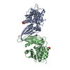 7zgtC  7zkgC 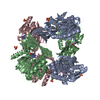 1wznS S: Starting model for refinement C: citing same article ( |
|---|---|
| Similar structure data |
-Links
 PDBj
PDBj

- Assembly
| Deposited unit | 
| ||||||||||||
|---|---|---|---|---|---|---|---|---|---|---|---|---|---|
| 1 | 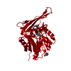
| ||||||||||||
| Unit cell |
| ||||||||||||
| Components on special symmetry positions |
|
-Components
| #1: Protein | Mass: 30780.125 Da / Num. of mol.: 1 Source method: isolated from a genetically manipulated source Source: (gene. exp.) Streptomyces griseofuscus (bacteria) / Gene: psmD / Production host: |
|---|---|
| #2: Chemical | ChemComp-UNL / Num. of mol.: 1 / Source method: obtained synthetically |
| #3: Chemical | ChemComp-PGE /   Mass: 150.173 Da / Num. of mol.: 1 / Source method: obtained synthetically / Formula: C6H14O4 Mass: 150.173 Da / Num. of mol.: 1 / Source method: obtained synthetically / Formula: C6H14O4 |
| #4: Chemical | ChemComp-SAH /   Mass: 384.411 Da / Num. of mol.: 1 / Source method: obtained synthetically / Formula: C14H20N6O5S / Feature type: SUBJECT OF INVESTIGATION Mass: 384.411 Da / Num. of mol.: 1 / Source method: obtained synthetically / Formula: C14H20N6O5S / Feature type: SUBJECT OF INVESTIGATION |
| #5: Water | ChemComp-HOH /  Mass: 18.015 Da / Num. of mol.: 295 / Source method: isolated from a natural source / Formula: H2O Mass: 18.015 Da / Num. of mol.: 295 / Source method: isolated from a natural source / Formula: H2O |
| Has ligand of interest | Y |
| Has protein modification | N |
-Experimental details
-Experiment
| Experiment | Method: X-RAY DIFFRACTION / Number of used crystals: 1 |
|---|
- Sample preparation
| Crystal | Density Matthews: 2.03 Å3/Da / Density % sol: 39.31 % |
|---|---|
| Crystal grow | Temperature: 293 K / Method: vapor diffusion, sitting drop / pH: 8.5 / Details: PEG 1000, Tris-HCl |
-Data collection
| Diffraction | Mean temperature: 100 K / Serial crystal experiment: N |
|---|---|
| Diffraction source | Source: SYNCHROTRON / Site: PETRA III, DESY  / Beamline: P11 / Wavelength: 1.0332 Å / Beamline: P11 / Wavelength: 1.0332 Å |
| Detector | Type: DECTRIS PILATUS 6M-F / Detector: PIXEL / Date: Jun 29, 2020 |
| Radiation | Protocol: SINGLE WAVELENGTH / Monochromatic (M) / Laue (L): M / Scattering type: x-ray |
| Radiation wavelength | Wavelength: 1.0332 Å / Relative weight: 1 |
| Reflection | Resolution: 1.4→53.36 Å / Num. obs: 49285 / % possible obs: 98.6 % / Redundancy: 8.2 % / Biso Wilson estimate: 21.39 Å2 / CC1/2: 1 / Rrim(I) all: 0.042 / Net I/σ(I): 22.73 |
| Reflection shell | Resolution: 1.4→1.44 Å / Redundancy: 5.94 % / Mean I/σ(I) obs: 0.93 / Num. unique obs: 3292 / CC1/2: 0.402 / Rrim(I) all: 2.005 / % possible all: 89.7 |
- Processing
| Software |
| |||||||||||||||||||||||||||||||||||||||||||||||||||||||||||||||||||||||||||||||||||||||||||||||||||||||||||||||||||||||||||||||||||||
|---|---|---|---|---|---|---|---|---|---|---|---|---|---|---|---|---|---|---|---|---|---|---|---|---|---|---|---|---|---|---|---|---|---|---|---|---|---|---|---|---|---|---|---|---|---|---|---|---|---|---|---|---|---|---|---|---|---|---|---|---|---|---|---|---|---|---|---|---|---|---|---|---|---|---|---|---|---|---|---|---|---|---|---|---|---|---|---|---|---|---|---|---|---|---|---|---|---|---|---|---|---|---|---|---|---|---|---|---|---|---|---|---|---|---|---|---|---|---|---|---|---|---|---|---|---|---|---|---|---|---|---|---|---|---|
| Refinement | Method to determine structure: MOLECULAR REPLACEMENT Starting model: 1WZN Resolution: 1.4→53.36 Å / SU ML: 0.1567 / Cross valid method: FREE R-VALUE / Phase error: 19.5096 Stereochemistry target values: GeoStd + Monomer Library + CDL v1.2
| |||||||||||||||||||||||||||||||||||||||||||||||||||||||||||||||||||||||||||||||||||||||||||||||||||||||||||||||||||||||||||||||||||||
| Solvent computation | Shrinkage radii: 0.9 Å / VDW probe radii: 1.11 Å / Solvent model: FLAT BULK SOLVENT MODEL | |||||||||||||||||||||||||||||||||||||||||||||||||||||||||||||||||||||||||||||||||||||||||||||||||||||||||||||||||||||||||||||||||||||
| Displacement parameters | Biso mean: 30.52 Å2 | |||||||||||||||||||||||||||||||||||||||||||||||||||||||||||||||||||||||||||||||||||||||||||||||||||||||||||||||||||||||||||||||||||||
| Refinement step | Cycle: LAST / Resolution: 1.4→53.36 Å
| |||||||||||||||||||||||||||||||||||||||||||||||||||||||||||||||||||||||||||||||||||||||||||||||||||||||||||||||||||||||||||||||||||||
| Refine LS restraints |
| |||||||||||||||||||||||||||||||||||||||||||||||||||||||||||||||||||||||||||||||||||||||||||||||||||||||||||||||||||||||||||||||||||||
| LS refinement shell |
|
