 Movie
Movie Controller

[English] 日本語
 Yorodumi
- PDB-7zjv: Structure of human USPL1 in covalent complex with DeltaN-SUMO2/3-... -
+ Open data

- Basic information
| Entry | Database: PDB / ID: 7zjv | ||||||
|---|---|---|---|---|---|---|---|
| Title | Structure of human USPL1 in covalent complex with DeltaN-SUMO2/3-PA probe | ||||||
 Components Components |
| ||||||
 Keywords Keywords | HYDROLASE / USP / SUMO / Ubiquitin / probe / ubiquitin-like modifier | ||||||
| Function / homology |  Function and homology information Function and homology informationsnRNA transcription / Cajal body organization / deSUMOylase activity / protein desumoylation / SUMO is proteolytically processed / SUMO is conjugated to E1 (UBA2:SAE1) / SUMO is transferred from E1 to E2 (UBE2I, UBC9) / Vitamin D (calciferol) metabolism / SUMO binding / SUMOylation of SUMOylation proteins ...snRNA transcription / Cajal body organization / deSUMOylase activity / protein desumoylation / SUMO is proteolytically processed / SUMO is conjugated to E1 (UBA2:SAE1) / SUMO is transferred from E1 to E2 (UBE2I, UBC9) / Vitamin D (calciferol) metabolism / SUMO binding / SUMOylation of SUMOylation proteins / SUMOylation of RNA binding proteins / SUMO transferase activity / SUMOylation of transcription factors / SUMOylation of DNA replication proteins / ubiquitin-like protein ligase binding / protein sumoylation / postsynaptic cytosol / Cajal body / SUMOylation of DNA damage response and repair proteins / presynaptic cytosol / SUMOylation of transcription cofactors / SUMOylation of chromatin organization proteins / hippocampal mossy fiber to CA3 synapse / Regulation of endogenous retroelements by KRAB-ZFP proteins / SUMOylation of intracellular receptors / PML body / GABA-ergic synapse / Formation of Incision Complex in GG-NER / protein tag activity / positive regulation of proteasomal ubiquitin-dependent protein catabolic process / Processing of DNA double-strand break ends / Hydrolases; Acting on peptide bonds (peptidases); Cysteine endopeptidases / cell population proliferation / ubiquitin protein ligase binding / glutamatergic synapse / positive regulation of transcription by RNA polymerase II / proteolysis / : / RNA binding / nucleoplasm / nucleus Similarity search - Function | ||||||
| Biological species |  Homo sapiens (human) Homo sapiens (human) | ||||||
| Method |  X-RAY DIFFRACTION / SYNCHROTRON / MOLECULAR REPLACEMENT / Resolution: 2.4 Å X-RAY DIFFRACTION / SYNCHROTRON / MOLECULAR REPLACEMENT / Resolution: 2.4 Å | ||||||
 Authors Authors | Zhao, Z. / Gersch, M. | ||||||
| Funding support |  Germany, 1items Germany, 1items
| ||||||
 Citation Citation | Journal: J.Am.Chem.Soc. / Year: 2023 Title: Native Semisynthesis of Isopeptide-Linked Substrates for Specificity Analysis of Deubiquitinases and Ubl Proteases. Authors: Zhao, Z. / O'Dea, R. / Wendrich, K. / Kazi, N. / Gersch, M. #1: Journal: Acta Crystallogr D Struct Biol / Year: 2019 Title: Macromolecular structure determination using X-rays, neutrons and electrons: recent developments in Phenix. Authors: Dorothee Liebschner / Pavel V Afonine / Matthew L Baker / Gábor Bunkóczi / Vincent B Chen / Tristan I Croll / Bradley Hintze / Li Wei Hung / Swati Jain / Airlie J McCoy / Nigel W Moriarty ...Authors: Dorothee Liebschner / Pavel V Afonine / Matthew L Baker / Gábor Bunkóczi / Vincent B Chen / Tristan I Croll / Bradley Hintze / Li Wei Hung / Swati Jain / Airlie J McCoy / Nigel W Moriarty / Robert D Oeffner / Billy K Poon / Michael G Prisant / Randy J Read / Jane S Richardson / David C Richardson / Massimo D Sammito / Oleg V Sobolev / Duncan H Stockwell / Thomas C Terwilliger / Alexandre G Urzhumtsev / Lizbeth L Videau / Christopher J Williams / Paul D Adams /    Abstract: Diffraction (X-ray, neutron and electron) and electron cryo-microscopy are powerful methods to determine three-dimensional macromolecular structures, which are required to understand biological ...Diffraction (X-ray, neutron and electron) and electron cryo-microscopy are powerful methods to determine three-dimensional macromolecular structures, which are required to understand biological processes and to develop new therapeutics against diseases. The overall structure-solution workflow is similar for these techniques, but nuances exist because the properties of the reduced experimental data are different. Software tools for structure determination should therefore be tailored for each method. Phenix is a comprehensive software package for macromolecular structure determination that handles data from any of these techniques. Tasks performed with Phenix include data-quality assessment, map improvement, model building, the validation/rebuilding/refinement cycle and deposition. Each tool caters to the type of experimental data. The design of Phenix emphasizes the automation of procedures, where possible, to minimize repetitive and time-consuming manual tasks, while default parameters are chosen to encourage best practice. A graphical user interface provides access to many command-line features of Phenix and streamlines the transition between programs, project tracking and re-running of previous tasks. | ||||||
| History |
|
- Structure visualization
| Structure viewer | Molecule: MolmilJmol/JSmol |
|---|
- Downloads & links
-Download
| PDBx/mmCIF format | 7zjv.cif.gz | 181.9 KB | Display | PDBx/mmCIF format |
|---|---|---|---|---|
| PDB format | pdb7zjv.ent.gz | 120.4 KB | Display | PDB format |
| PDBx/mmJSON format | 7zjv.json.gz | Tree view | PDBx/mmJSON format | |
| Others |  Other downloads Other downloads |
-Validation report
| Arichive directory | https://data.pdbj.org/pub/pdb/validation_reports/zj/7zjvftp://data.pdbj.org/pub/pdb/validation_reports/zj/7zjv | HTTPS FTP |
|---|
-Related structure data
| Related structure data | 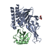 7zjuSC S: Starting model for refinement C: citing same article ( |
|---|---|
| Similar structure data |
-Links
 PDBj
PDBj


- Assembly
| Deposited unit | 
| ||||||||||||
|---|---|---|---|---|---|---|---|---|---|---|---|---|---|
| 1 |
| ||||||||||||
| Unit cell |
|
-Components
-Protein , 2 types, 2 molecules AB
| #1: Protein | Mass: 33203.062 Da / Num. of mol.: 1 Source method: isolated from a genetically manipulated source Source: (gene. exp.) Homo sapiens (human) / Gene: USPL1 / Production host:  |
|---|---|
| #2: Protein | Mass: 8769.975 Da / Num. of mol.: 1 Source method: isolated from a genetically manipulated source Source: (gene. exp.) Homo sapiens (human) / Gene: SUMO2, SMT3B, SMT3H2 / Production host: |
-Non-polymers , 4 types, 35 molecules 


| #3: Chemical | ChemComp-ZN /  Mass: 65.409 Da / Num. of mol.: 1 / Source method: obtained synthetically / Formula: Zn Mass: 65.409 Da / Num. of mol.: 1 / Source method: obtained synthetically / Formula: Zn | ||
|---|---|---|---|
| #4: Chemical | ChemComp-AYE /  Mass: 57.094 Da / Num. of mol.: 1 / Source method: obtained synthetically / Formula: C3H7N / Feature type: SUBJECT OF INVESTIGATION Mass: 57.094 Da / Num. of mol.: 1 / Source method: obtained synthetically / Formula: C3H7N / Feature type: SUBJECT OF INVESTIGATION | ||
| #5: Chemical | ChemComp-CL /  Mass: 35.453 Da / Num. of mol.: 5 / Source method: obtained synthetically / Formula: Cl Mass: 35.453 Da / Num. of mol.: 5 / Source method: obtained synthetically / Formula: Cl#6: Water | ChemComp-HOH / | Mass: 18.015 Da / Num. of mol.: 28 / Source method: isolated from a natural source / Formula: H2O |
-Details
| Has ligand of interest | Y |
|---|---|
| Has protein modification | Y |
-Experimental details
-Experiment
| Experiment | Method: X-RAY DIFFRACTION / Number of used crystals: 1 |
|---|
- Sample preparation
| Crystal | Density Matthews: 2.26 Å3/Da / Density % sol: 45.6 % |
|---|---|
| Crystal grow | Temperature: 293 K / Method: vapor diffusion, sitting drop / pH: 8.9 / Details: 100 mM CHES pH 8.9, 34% PEG600 |
-Data collection
| Diffraction | Mean temperature: 100 K / Serial crystal experiment: N |
|---|---|
| Diffraction source | Source: SYNCHROTRON / Site: SLS  / Beamline: X10SA / Wavelength: 1 Å / Beamline: X10SA / Wavelength: 1 Å |
| Detector | Type: DECTRIS EIGER2 X 16M / Detector: PIXEL / Date: Jun 22, 2020 |
| Radiation | Protocol: SINGLE WAVELENGTH / Monochromatic (M) / Laue (L): M / Scattering type: x-ray |
| Radiation wavelength | Wavelength: 1 Å / Relative weight: 1 |
| Reflection | Resolution: 2.4→42.8 Å / Num. obs: 28746 / % possible obs: 100 % / Redundancy: 9.1 % / Biso Wilson estimate: 76.34 Å2 / CC1/2: 0.999 / Rmerge(I) obs: 0.053 / Rrim(I) all: 0.057 / Net I/σ(I): 18.2 |
| Reflection shell | Resolution: 2.4→2.49 Å / Rmerge(I) obs: 0.858 / Mean I/σ(I) obs: 1.8 / Num. unique obs: 1597 / CC1/2: 0.718 / Rrim(I) all: 0.933 |
- Processing
| Software |
| |||||||||||||||||||||||||||||||||||||||||||||||||||||||||||||||||||||||||||||||||||||||||||||||||||||||||||||||||||||||||||||||||||||||||||||||||||||||||||||||||||||||||||||||
|---|---|---|---|---|---|---|---|---|---|---|---|---|---|---|---|---|---|---|---|---|---|---|---|---|---|---|---|---|---|---|---|---|---|---|---|---|---|---|---|---|---|---|---|---|---|---|---|---|---|---|---|---|---|---|---|---|---|---|---|---|---|---|---|---|---|---|---|---|---|---|---|---|---|---|---|---|---|---|---|---|---|---|---|---|---|---|---|---|---|---|---|---|---|---|---|---|---|---|---|---|---|---|---|---|---|---|---|---|---|---|---|---|---|---|---|---|---|---|---|---|---|---|---|---|---|---|---|---|---|---|---|---|---|---|---|---|---|---|---|---|---|---|---|---|---|---|---|---|---|---|---|---|---|---|---|---|---|---|---|---|---|---|---|---|---|---|---|---|---|---|---|---|---|---|---|---|
| Refinement | Method to determine structure: MOLECULAR REPLACEMENT Starting model: 7ZJU Resolution: 2.4→42.8 Å / SU ML: 0.4606 / Cross valid method: FREE R-VALUE / σ(F): 1.34 / Phase error: 35.2751 Stereochemistry target values: GeoStd + Monomer Library + CDL v1.2
| |||||||||||||||||||||||||||||||||||||||||||||||||||||||||||||||||||||||||||||||||||||||||||||||||||||||||||||||||||||||||||||||||||||||||||||||||||||||||||||||||||||||||||||||
| Solvent computation | Shrinkage radii: 0.9 Å / VDW probe radii: 1.11 Å / Solvent model: FLAT BULK SOLVENT MODEL | |||||||||||||||||||||||||||||||||||||||||||||||||||||||||||||||||||||||||||||||||||||||||||||||||||||||||||||||||||||||||||||||||||||||||||||||||||||||||||||||||||||||||||||||
| Displacement parameters | Biso mean: 109.6 Å2 | |||||||||||||||||||||||||||||||||||||||||||||||||||||||||||||||||||||||||||||||||||||||||||||||||||||||||||||||||||||||||||||||||||||||||||||||||||||||||||||||||||||||||||||||
| Refinement step | Cycle: LAST / Resolution: 2.4→42.8 Å
| |||||||||||||||||||||||||||||||||||||||||||||||||||||||||||||||||||||||||||||||||||||||||||||||||||||||||||||||||||||||||||||||||||||||||||||||||||||||||||||||||||||||||||||||
| Refine LS restraints |
| |||||||||||||||||||||||||||||||||||||||||||||||||||||||||||||||||||||||||||||||||||||||||||||||||||||||||||||||||||||||||||||||||||||||||||||||||||||||||||||||||||||||||||||||
| LS refinement shell |
| |||||||||||||||||||||||||||||||||||||||||||||||||||||||||||||||||||||||||||||||||||||||||||||||||||||||||||||||||||||||||||||||||||||||||||||||||||||||||||||||||||||||||||||||
| Refinement TLS params. | Method: refined / Refine-ID: X-RAY DIFFRACTION
| |||||||||||||||||||||||||||||||||||||||||||||||||||||||||||||||||||||||||||||||||||||||||||||||||||||||||||||||||||||||||||||||||||||||||||||||||||||||||||||||||||||||||||||||
| Refinement TLS group | Refine-ID: X-RAY DIFFRACTION
|
