 Movie
Movie Controller


+ Open data

- Basic information
| Entry | Database: PDB / ID: 7zir | ||||||||||||
|---|---|---|---|---|---|---|---|---|---|---|---|---|---|
| Title | Cryo-EM structure of hnRNPDL amyloid fibrils | ||||||||||||
 Components Components | Heterogeneous nuclear ribonucleoprotein D-like | ||||||||||||
 Keywords Keywords | RNA BINDING PROTEIN / Amyloid / Protein aggregation / Alternative splicing / Exon / Prion-like / LGMD1G / cryoEM structure | ||||||||||||
| Function / homology |  Function and homology information Function and homology informationpoly(G) binding / poly(A) binding / RNA processing / Gene and protein expression by JAK-STAT signaling after Interleukin-12 stimulation / spliceosomal complex / single-stranded DNA binding / regulation of gene expression / double-stranded DNA binding / chromatin / DNA binding ...poly(G) binding / poly(A) binding / RNA processing / Gene and protein expression by JAK-STAT signaling after Interleukin-12 stimulation / spliceosomal complex / single-stranded DNA binding / regulation of gene expression / double-stranded DNA binding / chromatin / DNA binding / RNA binding / nucleoplasm / nucleus / cytosol Similarity search - Function | ||||||||||||
| Biological species |  Homo sapiens (human) Homo sapiens (human) | ||||||||||||
| Method | ELECTRON MICROSCOPY / helical reconstruction / cryo EM / Resolution: 2.5 Å | ||||||||||||
 Authors Authors | Garcia-Pardo, J. / Chaves-Sanjuan, A. / Bartolome-Nafria, A. / Gil-Garcia, M. / Visentin, C. / Bolognesi, M. / Ricagno, S. / Ventura, S. | ||||||||||||
| Funding support |  Spain, 3items Spain, 3items
| ||||||||||||
 Citation Citation | Journal: Nat Commun / Year: 2023 Title: Cryo-EM structure of hnRNPDL-2 fibrils, a functional amyloid associated with limb-girdle muscular dystrophy D3. Authors: Javier Garcia-Pardo / Andrea Bartolomé-Nafría / Antonio Chaves-Sanjuan / Marcos Gil-Garcia / Cristina Visentin / Martino Bolognesi / Stefano Ricagno / Salvador Ventura /  Abstract: hnRNPDL is a ribonucleoprotein (RNP) involved in transcription and RNA-processing that hosts missense mutations causing limb-girdle muscular dystrophy D3 (LGMD D3). Mammalian-specific alternative ...hnRNPDL is a ribonucleoprotein (RNP) involved in transcription and RNA-processing that hosts missense mutations causing limb-girdle muscular dystrophy D3 (LGMD D3). Mammalian-specific alternative splicing (AS) renders three natural isoforms, hnRNPDL-2 being predominant in humans. We present the cryo-electron microscopy structure of full-length hnRNPDL-2 amyloid fibrils, which are stable, non-toxic, and bind nucleic acids. The high-resolution amyloid core consists of a single Gly/Tyr-rich and highly hydrophilic filament containing internal water channels. The RNA binding domains are located as a solenoidal coat around the core. The architecture and activity of hnRNPDL-2 fibrils are reminiscent of functional amyloids, our results suggesting that LGMD D3 might be a loss-of-function disease associated with impaired fibrillation. Strikingly, the fibril core matches exon 6, absent in the soluble hnRNPDL-3 isoform. This provides structural evidence for AS controlling hnRNPDL assembly by precisely including/skipping an amyloid exon, a mechanism that holds the potential to generate functional diversity in RNPs. | ||||||||||||
| History |
|
- Structure visualization
| Structure viewer | Molecule: MolmilJmol/JSmol |
|---|
- Downloads & links
-Download
| PDBx/mmCIF format | 7zir.cif.gz | 88.2 KB | Display | PDBx/mmCIF format |
|---|---|---|---|---|
| PDB format | pdb7zir.ent.gz | 50.2 KB | Display | PDB format |
| PDBx/mmJSON format | 7zir.json.gz | Tree view | PDBx/mmJSON format | |
| Others |  Other downloads Other downloads |
-Validation report
| Summary document | 7zir_validation.pdf.gz | 967.7 KB | Display | wwPDB validaton report |
|---|---|---|---|---|
| Full document | 7zir_full_validation.pdf.gz | 968 KB | Display | |
| Data in XML | 7zir_validation.xml.gz | 27.5 KB | Display | |
| Data in CIF | 7zir_validation.cif.gz | 38.1 KB | Display | |
| Arichive directory | https://data.pdbj.org/pub/pdb/validation_reports/zi/7zirftp://data.pdbj.org/pub/pdb/validation_reports/zi/7zir | HTTPS FTP |
-Related structure data
| Related structure data |  14738MC M: map data used to model this data C: citing same article ( |
|---|---|
| Similar structure data |
-Links
 PDBj
PDBj


- Assembly
| Deposited unit | 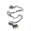
|
|---|---|
| 1 |
|
-Components
| #1: Protein | Mass: 45804.266 Da / Num. of mol.: 5 Source method: isolated from a genetically manipulated source Source: (gene. exp.) Homo sapiens (human) / Gene: HNRNPDL, HNRPDL, JKTBP / Production host:  #2: Water | ChemComp-HOH / |  Mass: 18.015 Da / Num. of mol.: 50 / Source method: isolated from a natural source / Formula: H2O Mass: 18.015 Da / Num. of mol.: 50 / Source method: isolated from a natural source / Formula: H2O |
|---|
-Experimental details
-Experiment
| Experiment | Method: ELECTRON MICROSCOPY |
|---|---|
| EM experiment | Aggregation state: FILAMENT / 3D reconstruction method: helical reconstruction |
- Sample preparation
| Component | Name: hnRNPDL-2 amyloid fibrils / Type: COMPLEX Details: Fibril structure of the recombinantly produced full-lenght Heterogeneous ribonucleoprotein D-like (hnRNPDL) isoform 2 Entity ID: #1 / Source: RECOMBINANT | ||||||||||||
|---|---|---|---|---|---|---|---|---|---|---|---|---|---|
| Molecular weight | Experimental value: NO | ||||||||||||
| Source (natural) | Organism: Homo sapiens (human) | ||||||||||||
| Source (recombinant) | Organism: | ||||||||||||
| Buffer solution | pH: 7.5 | ||||||||||||
| Buffer component |
| ||||||||||||
| Specimen | Conc.: 0.23 mg/ml / Embedding applied: NO / Shadowing applied: NO / Staining applied: NO / Vitrification applied: YES Details: Diluted sample of hnRNPDL2 amyloid fibrils formed under native conditions. | ||||||||||||
| Specimen support | Grid material: COPPER / Grid mesh size: 300 divisions/in. / Grid type: C-flat-1.2/1.3 | ||||||||||||
| Vitrification | Instrument: FEI VITROBOT MARK IV / Cryogen name: ETHANE / Humidity: 100 % |
- Electron microscopy imaging
| Experimental equipment |  Model: Talos Arctica / Image courtesy: FEI Company |
|---|---|
| Microscopy | Model: FEI TALOS ARCTICA |
| Electron gun | Electron source:  FIELD EMISSION GUN / Accelerating voltage: 200 kV / Illumination mode: FLOOD BEAM FIELD EMISSION GUN / Accelerating voltage: 200 kV / Illumination mode: FLOOD BEAM |
| Electron lens | Mode: BRIGHT FIELD / Nominal magnification: 120000 X / Nominal defocus max: 2200 nm / Nominal defocus min: 1000 nm / Cs: 2.7 mm / C2 aperture diameter: 50 µm / Alignment procedure: COMA FREE |
| Specimen holder | Cryogen: NITROGEN |
| Image recording | Electron dose: 40 e/Å2 / Film or detector model: FEI FALCON III (4k x 4k) / Num. of grids imaged: 1 / Num. of real images: 1114 |
- Processing
| Software | Name: PHENIX / Version: 1.19.2_4158: / Classification: refinement | ||||||||||||||||||||||||||||
|---|---|---|---|---|---|---|---|---|---|---|---|---|---|---|---|---|---|---|---|---|---|---|---|---|---|---|---|---|---|
| EM software |
| ||||||||||||||||||||||||||||
| CTF correction | Type: NONE | ||||||||||||||||||||||||||||
| Helical symmerty | Angular rotation/subunit: -4.8 ° / Axial rise/subunit: 4.8 Å / Axial symmetry: C1 | ||||||||||||||||||||||||||||
| Particle selection | Num. of particles selected: 158493 | ||||||||||||||||||||||||||||
| 3D reconstruction | Resolution: 2.5 Å / Resolution method: FSC 0.143 CUT-OFF / Num. of particles: 54490 / Num. of class averages: 1 / Symmetry type: HELICAL | ||||||||||||||||||||||||||||
| Atomic model building | Protocol: BACKBONE TRACE / Space: REAL | ||||||||||||||||||||||||||||
| Refine LS restraints |
|
