 Movie
Movie Controller

[English] 日本語
 Yorodumi
- PDB-7zii: Crystal structure of human tryptophan hydroxylase 1 in complex wi... -
+ Open data

- Basic information
| Entry | Database: PDB / ID: 7zii | ||||||
|---|---|---|---|---|---|---|---|
| Title | Crystal structure of human tryptophan hydroxylase 1 in complex with inhibitor KM-05-193 | ||||||
 Components Components | Tryptophan 5-hydroxylase 1 | ||||||
 Keywords Keywords | METAL BINDING PROTEIN / tryptophan hydroxylase / serotonin biosynthesis | ||||||
| Function / homology |  Function and homology information Function and homology informationregulation of hemostasis / : / tryptophan 5-monooxygenase / tryptophan 5-monooxygenase activity / Serotonin and melatonin biosynthesis / serotonin biosynthetic process / platelet degranulation / NGF-stimulated transcription / bone remodeling / mammary gland alveolus development ...regulation of hemostasis / : / tryptophan 5-monooxygenase / tryptophan 5-monooxygenase activity / Serotonin and melatonin biosynthesis / serotonin biosynthetic process / platelet degranulation / NGF-stimulated transcription / bone remodeling / mammary gland alveolus development / positive regulation of fat cell differentiation / neuron projection / iron ion binding / cytosol Similarity search - Function | ||||||
| Biological species |  Homo sapiens (human) Homo sapiens (human) | ||||||
| Method |  X-RAY DIFFRACTION / SYNCHROTRON / MOLECULAR REPLACEMENT / Resolution: 1.62800050605 Å X-RAY DIFFRACTION / SYNCHROTRON / MOLECULAR REPLACEMENT / Resolution: 1.62800050605 Å | ||||||
 Authors Authors | Schuetz, A. / Mallow, K. / Nazare, M. / Specker, E. / Heinemann, U. | ||||||
| Funding support |  Germany, 1items Germany, 1items
| ||||||
 Citation Citation | Journal: J.Med.Chem. / Year: 2022 Title: Structure-Based Design of Xanthine-Benzimidazole Derivatives as Novel and Potent Tryptophan Hydroxylase Inhibitors. Authors: Specker, E. / Matthes, S. / Wesolowski, R. / Schutz, A. / Grohmann, M. / Alenina, N. / Pleimes, D. / Mallow, K. / Neuenschwander, M. / Gogolin, A. / Weise, M. / Pfeifer, J. / Ziebart, N. / ...Authors: Specker, E. / Matthes, S. / Wesolowski, R. / Schutz, A. / Grohmann, M. / Alenina, N. / Pleimes, D. / Mallow, K. / Neuenschwander, M. / Gogolin, A. / Weise, M. / Pfeifer, J. / Ziebart, N. / Heinemann, U. / von Kries, J.P. / Nazare, M. / Bader, M. | ||||||
| History |
|
- Structure visualization
| Structure viewer | Molecule: MolmilJmol/JSmol |
|---|
- Downloads & links
-Download
| PDBx/mmCIF format | 7zii.cif.gz | 100.3 KB | Display | PDBx/mmCIF format |
|---|---|---|---|---|
| PDB format | pdb7zii.ent.gz | 58.8 KB | Display | PDB format |
| PDBx/mmJSON format | 7zii.json.gz | Tree view | PDBx/mmJSON format | |
| Others |  Other downloads Other downloads |
-Validation report
| Arichive directory | https://data.pdbj.org/pub/pdb/validation_reports/zi/7ziiftp://data.pdbj.org/pub/pdb/validation_reports/zi/7zii | HTTPS FTP |
|---|
-Related structure data
| Related structure data | 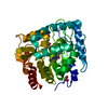 7zifC  7zigC 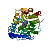 7zihC  7zijC 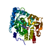 1mlwS S: Starting model for refinement C: citing same article ( |
|---|---|
| Similar structure data |
-Links
 PDBj
PDBj


- Assembly
| Deposited unit | 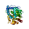
| ||||||||||||
|---|---|---|---|---|---|---|---|---|---|---|---|---|---|
| 1 |
| ||||||||||||
| Unit cell |
|
-Components
| #1: Protein | Mass: 37424.602 Da / Num. of mol.: 1 Source method: isolated from a genetically manipulated source Details: catalytic domain of human TPH1 / Source: (gene. exp.) Homo sapiens (human) / Gene: TPH1, TPH, TPRH, TRPH / Production host:  |
|---|---|
| #2: Chemical | ChemComp-FE /   Mass: 55.845 Da / Num. of mol.: 1 / Source method: obtained synthetically / Formula: Fe Mass: 55.845 Da / Num. of mol.: 1 / Source method: obtained synthetically / Formula: Fe |
| #3: Chemical | ChemComp-IVQ / 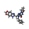  Mass: 458.469 Da / Num. of mol.: 1 / Source method: obtained synthetically / Formula: C24H22N6O4 / Feature type: SUBJECT OF INVESTIGATION Mass: 458.469 Da / Num. of mol.: 1 / Source method: obtained synthetically / Formula: C24H22N6O4 / Feature type: SUBJECT OF INVESTIGATION |
| #4: Chemical | ChemComp-GOL /   Mass: 92.094 Da / Num. of mol.: 1 / Source method: obtained synthetically / Formula: C3H8O3 Mass: 92.094 Da / Num. of mol.: 1 / Source method: obtained synthetically / Formula: C3H8O3 |
| #5: Water | ChemComp-HOH /  Mass: 18.015 Da / Num. of mol.: 314 / Source method: isolated from a natural source / Formula: H2O Mass: 18.015 Da / Num. of mol.: 314 / Source method: isolated from a natural source / Formula: H2O |
| Has ligand of interest | Y |
-Experimental details
-Experiment
| Experiment | Method: X-RAY DIFFRACTION / Number of used crystals: 1 |
|---|
- Sample preparation
| Crystal | Density Matthews: 2.14 Å3/Da / Density % sol: 42.45 % |
|---|---|
| Crystal grow | Temperature: 293.15 K / Method: vapor diffusion, sitting drop / Details: 20% w/v PEG 3350, 200 mM potassium formate pH 7.3 |
-Data collection
| Diffraction | Mean temperature: 100 K / Serial crystal experiment: N |
|---|---|
| Diffraction source | Source: SYNCHROTRON / Site: BESSY / Beamline: 14.1 / Wavelength: 0.9184 Å |
| Detector | Type: DECTRIS PILATUS 6M / Detector: PIXEL / Date: Jul 19, 2015 |
| Radiation | Protocol: SINGLE WAVELENGTH / Monochromatic (M) / Laue (L): M / Scattering type: x-ray |
| Radiation wavelength | Wavelength: 0.9184 Å / Relative weight: 1 |
| Reflection | Resolution: 1.628→29.5515963785 Å / Num. obs: 39546 / % possible obs: 99.66 % / Redundancy: 4.5 % / Biso Wilson estimate: 17.8251303948 Å2 / Rmerge(I) obs: 0.0669 / Net I/σ(I): 14.71 |
| Reflection shell | Resolution: 1.63→1.69 Å / Rmerge(I) obs: 0.7235 / Num. unique obs: 3859 |
- Processing
| Software |
| ||||||||||||||||||||||||||||||||||||||||||||||||||||||||||||||||||||||||||||||||||||||||||||||||||
|---|---|---|---|---|---|---|---|---|---|---|---|---|---|---|---|---|---|---|---|---|---|---|---|---|---|---|---|---|---|---|---|---|---|---|---|---|---|---|---|---|---|---|---|---|---|---|---|---|---|---|---|---|---|---|---|---|---|---|---|---|---|---|---|---|---|---|---|---|---|---|---|---|---|---|---|---|---|---|---|---|---|---|---|---|---|---|---|---|---|---|---|---|---|---|---|---|---|---|---|
| Refinement | Method to determine structure: MOLECULAR REPLACEMENT Starting model: 1mlw Resolution: 1.62800050605→29.5515963785 Å / SU ML: 0.169431605722 / Cross valid method: FREE R-VALUE / σ(F): 1.3544379811 / Phase error: 20.1215586376
| ||||||||||||||||||||||||||||||||||||||||||||||||||||||||||||||||||||||||||||||||||||||||||||||||||
| Solvent computation | Shrinkage radii: 0.9 Å / VDW probe radii: 1.11 Å / Solvent model: FLAT BULK SOLVENT MODEL | ||||||||||||||||||||||||||||||||||||||||||||||||||||||||||||||||||||||||||||||||||||||||||||||||||
| Displacement parameters | Biso mean: 25.8363252539 Å2 | ||||||||||||||||||||||||||||||||||||||||||||||||||||||||||||||||||||||||||||||||||||||||||||||||||
| Refinement step | Cycle: LAST / Resolution: 1.62800050605→29.5515963785 Å
| ||||||||||||||||||||||||||||||||||||||||||||||||||||||||||||||||||||||||||||||||||||||||||||||||||
| Refine LS restraints |
| ||||||||||||||||||||||||||||||||||||||||||||||||||||||||||||||||||||||||||||||||||||||||||||||||||
| LS refinement shell |
|
