 Movie
Movie Controller


+ Open data

- Basic information
| Entry | Database: PDB / ID: 7zhd | ||||||
|---|---|---|---|---|---|---|---|
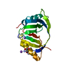
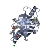
| Title | Crystal structure of CtaZ in complex with Closthioamide | ||||||
 Components Components | Transcription activator effector binding | ||||||
 Keywords Keywords | LIPID BINDING PROTEIN / GYRASE-LIKE DOMAIN / RECEPTOR / SIDEROPHORE / SELF PROTECTION / ANTIBIOTIC RESISTANCE / DRUG BINDING | ||||||
| Function / homology |  Function and homology information Function and homology information | ||||||
| Biological species |  Ruminiclostridium cellulolyticum (bacteria) Ruminiclostridium cellulolyticum (bacteria) | ||||||
| Method |  X-RAY DIFFRACTION / SYNCHROTRON / SAD / Resolution: 1.65 Å X-RAY DIFFRACTION / SYNCHROTRON / SAD / Resolution: 1.65 Å | ||||||
 Authors Authors | Gude, F. / Molloy, E.M. / Horch, T. / Dell, M. / Dunbar, K.L. / Krabbe, J. / Groll, M. / Hertweck, C. | ||||||
| Funding support | European Union, 1items
| ||||||
 Citation Citation | Journal: Angew.Chem.Int.Ed.Engl. / Year: 2022 Title: A Specialized Polythioamide-Binding Protein Confers Antibiotic Self-Resistance in Anaerobic Bacteria. Authors: Gude, F. / Molloy, E.M. / Horch, T. / Dell, M. / Dunbar, K.L. / Krabbe, J. / Groll, M. / Hertweck, C. | ||||||
| History |
|
- Structure visualization
| Structure viewer | Molecule: MolmilJmol/JSmol |
|---|
- Downloads & links
-Download
| PDBx/mmCIF format | 7zhd.cif.gz | 80.5 KB | Display | PDBx/mmCIF format |
|---|---|---|---|---|
| PDB format | pdb7zhd.ent.gz | 58.6 KB | Display | PDB format |
| PDBx/mmJSON format | 7zhd.json.gz | Tree view | PDBx/mmJSON format | |
| Others |  Other downloads Other downloads |
-Validation report
| Summary document | 7zhd_validation.pdf.gz | 663.8 KB | Display | wwPDB validaton report |
|---|---|---|---|---|
| Full document | 7zhd_full_validation.pdf.gz | 664.7 KB | Display | |
| Data in XML | 7zhd_validation.xml.gz | 8.9 KB | Display | |
| Data in CIF | 7zhd_validation.cif.gz | 11.5 KB | Display | |
| Arichive directory | https://data.pdbj.org/pub/pdb/validation_reports/zh/7zhdftp://data.pdbj.org/pub/pdb/validation_reports/zh/7zhd | HTTPS FTP |
-Related structure data
-Links
 PDBj
PDBj- Assembly
| Deposited unit | 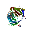
| ||||||||
|---|---|---|---|---|---|---|---|---|---|
| 1 |
| ||||||||
| Unit cell |
|
-Components
-Protein , 1 types, 1 molecules A
| #1: Protein | Mass: 17447.268 Da / Num. of mol.: 1 Source method: isolated from a genetically manipulated source Source: (gene. exp.) Ruminiclostridium cellulolyticum (bacteria)Gene: Ccel_3263 / Plasmid: pET28a / Production host: |
|---|
-Non-polymers , 5 types, 70 molecules 


| #2: Chemical | ChemComp-IQ4 / ~{ Mass: 695.041 Da / Num. of mol.: 1 / Source method: obtained synthetically / Formula: C29H38N6O2S6 / Feature type: SUBJECT OF INVESTIGATION Mass: 695.041 Da / Num. of mol.: 1 / Source method: obtained synthetically / Formula: C29H38N6O2S6 / Feature type: SUBJECT OF INVESTIGATION | ||||||
|---|---|---|---|---|---|---|---|
| #3: Chemical |  Mass: 65.409 Da / Num. of mol.: 2 / Source method: obtained synthetically / Formula: Zn Mass: 65.409 Da / Num. of mol.: 2 / Source method: obtained synthetically / Formula: Zn#4: Chemical | ChemComp-NA / |  Mass: 22.990 Da / Num. of mol.: 1 / Source method: obtained synthetically / Formula: Na Mass: 22.990 Da / Num. of mol.: 1 / Source method: obtained synthetically / Formula: Na#5: Chemical | ChemComp-TRS / |  Mass: 122.143 Da / Num. of mol.: 1 / Source method: obtained synthetically / Formula: C4H12NO3 / Comment: pH buffer*YM Mass: 122.143 Da / Num. of mol.: 1 / Source method: obtained synthetically / Formula: C4H12NO3 / Comment: pH buffer*YM#6: Water | ChemComp-HOH / | Mass: 18.015 Da / Num. of mol.: 65 / Source method: isolated from a natural source / Formula: H2O |
-Details
| Has ligand of interest | Y |
|---|
-Experimental details
-Experiment
| Experiment | Method: X-RAY DIFFRACTION / Number of used crystals: 1 |
|---|
- Sample preparation
| Crystal | Density Matthews: 2.74 Å3/Da / Density % sol: 55.17 % |
|---|---|
| Crystal grow | Temperature: 293 K / Method: vapor diffusion, sitting drop / pH: 6.5 / Details: 100 mM Na-cacodylate, 200 mM ZnAc2, 18% PEG 8000 |
-Data collection
| Diffraction | Mean temperature: 100 K / Serial crystal experiment: N |
|---|---|
| Diffraction source | Source: SYNCHROTRON / Site: SLS  / Beamline: X06SA / Wavelength: 1 Å / Beamline: X06SA / Wavelength: 1 Å |
| Detector | Type: DECTRIS EIGER X 16M / Detector: PIXEL / Date: Aug 29, 2020 |
| Radiation | Protocol: SINGLE WAVELENGTH / Monochromatic (M) / Laue (L): M / Scattering type: x-ray |
| Radiation wavelength | Wavelength: 1 Å / Relative weight: 1 |
| Reflection | Resolution: 1.65→50 Å / Num. obs: 22520 / % possible obs: 97.9 % / Redundancy: 3 % / Rmerge(I) obs: 0.033 / Net I/σ(I): 15.9 |
| Reflection shell | Resolution: 1.65→1.75 Å / Rmerge(I) obs: 0.548 / Mean I/σ(I) obs: 2.2 / Num. unique obs: 3648 / % possible all: 99.3 |
- Processing
| Software |
| |||||||||||||||||||||||||||||||||||||||||||||||||||||||||||||||||
|---|---|---|---|---|---|---|---|---|---|---|---|---|---|---|---|---|---|---|---|---|---|---|---|---|---|---|---|---|---|---|---|---|---|---|---|---|---|---|---|---|---|---|---|---|---|---|---|---|---|---|---|---|---|---|---|---|---|---|---|---|---|---|---|---|---|---|
| Refinement | Method to determine structure: SAD / Resolution: 1.65→30 Å / Cor.coef. Fo:Fc: 0.97 / Cor.coef. Fo:Fc free: 0.96 / SU B: 5.036 / SU ML: 0.072 / Cross valid method: THROUGHOUT / σ(F): 0 / ESU R: 0.106 / ESU R Free: 0.082 / Stereochemistry target values: MAXIMUM LIKELIHOOD Details: HYDROGENS HAVE BEEN ADDED IN THE RIDING POSITIONS U VALUES : WITH TLS ADDED
| |||||||||||||||||||||||||||||||||||||||||||||||||||||||||||||||||
| Solvent computation | Ion probe radii: 0.8 Å / Shrinkage radii: 0.8 Å / VDW probe radii: 1.2 Å / Solvent model: MASK | |||||||||||||||||||||||||||||||||||||||||||||||||||||||||||||||||
| Displacement parameters | Biso max: 76.09 Å2 / Biso mean: 35.699 Å2 / Biso min: 22.25 Å2
| |||||||||||||||||||||||||||||||||||||||||||||||||||||||||||||||||
| Refinement step | Cycle: final / Resolution: 1.65→30 Å
| |||||||||||||||||||||||||||||||||||||||||||||||||||||||||||||||||
| Refine LS restraints |
| |||||||||||||||||||||||||||||||||||||||||||||||||||||||||||||||||
| LS refinement shell | Resolution: 1.65→1.693 Å / Rfactor Rfree error: 0 / Total num. of bins used: 20
| |||||||||||||||||||||||||||||||||||||||||||||||||||||||||||||||||
| Refinement TLS params. | Method: refined / Origin x: 33.0722 Å / Origin y: 38.5759 Å / Origin z: 30.2599 Å
|
