- PDB-7zh7: Cryo-EM structure of ex vivo AA amyloid from renal tissue of a sh... -
+
Open data
ID or keywords:
Loading...
-
Basic information
Entry
Database: PDB / ID: 7zh7
Title
Cryo-EM structure of ex vivo AA amyloid from renal tissue of a short hair cat deceased in a shelter
Components
Serum amyloid A protein
Keywords
PROTEIN FIBRIL / AA amyloidosis / cat / Serum amyloid A
Function / homology
Serum amyloid A protein / : / Serum amyloid A protein / Serum amyloid A proteins signature. / Serum amyloid A proteins / high-density lipoprotein particle / acute-phase response / Serum amyloid A protein
Function and homology information
Biological species
Felis catus (domestic cat)
Method
ELECTRON MICROSCOPY / helical reconstruction / cryo EM / Resolution: 3.3 Å
Journal: Nat Commun / Year: 2022 Title: Cryo-EM structure of ex vivo fibrils associated with extreme AA amyloidosis prevalence in a cat shelter. Authors: Tim Schulte / Antonio Chaves-Sanjuan / Giulia Mazzini / Valentina Speranzini / Francesca Lavatelli / Filippo Ferri / Carlo Palizzotto / Maria Mazza / Paolo Milani / Mario Nuvolone / Anne- ...Authors: Tim Schulte / Antonio Chaves-Sanjuan / Giulia Mazzini / Valentina Speranzini / Francesca Lavatelli / Filippo Ferri / Carlo Palizzotto / Maria Mazza / Paolo Milani / Mario Nuvolone / Anne-Cathrine Vogt / Monique Vogel / Giovanni Palladini / Giampaolo Merlini / Martino Bolognesi / Silvia Ferro / Eric Zini / Stefano Ricagno / Abstract: AA amyloidosis is a systemic disease characterized by deposition of misfolded serum amyloid A protein (SAA) into cross-β amyloid in multiple organs in humans and animals. AA amyloidosis occurs at ...AA amyloidosis is a systemic disease characterized by deposition of misfolded serum amyloid A protein (SAA) into cross-β amyloid in multiple organs in humans and animals. AA amyloidosis occurs at high SAA serum levels during chronic inflammation. Prion-like transmission was reported as possible cause of extreme AA amyloidosis prevalence in captive animals, e.g. 70% in cheetah and 57-73% in domestic short hair (DSH) cats kept in zoos and shelters, respectively. Herein, we present the 3.3 Å cryo-EM structure of AA amyloid extracted post-mortem from the kidney of a DSH cat with renal failure, deceased in a shelter with extreme disease prevalence. The structure reveals a cross-β architecture assembled from two 76-residue long proto-filaments. Despite >70% sequence homology to mouse and human SAA, the cat SAA variant adopts a distinct amyloid fold. Inclusion of an eight-residue insert unique to feline SAA contributes to increased amyloid stability. The presented feline AA amyloid structure is fully compatible with the 99% identical amino acid sequence of amyloid fragments of captive cheetah.
A: Serum amyloid A protein B: Serum amyloid A protein C: Serum amyloid A protein D: Serum amyloid A protein E: Serum amyloid A protein F: Serum amyloid A protein G: Serum amyloid A protein H: Serum amyloid A protein I: Serum amyloid A protein J: Serum amyloid A protein
Embedding applied: NO / Shadowing applied: NO / Staining applied: NO / Vitrification applied: YES
Specimen support
Details: glow discharged for 30s at 30mA using a GloQube system Grid material: COPPER / Grid mesh size: 300 divisions/in. / Grid type: C-flat-1.2/1.3
Vitrification
Instrument: FEI VITROBOT MARK IV / Cryogen name: ETHANE / Humidity: 100 %
-
Electron microscopy imaging
Experimental equipment
Model: Talos Arctica / Image courtesy: FEI Company
Microscopy
Model: FEI TALOS ARCTICA
Electron gun
Electron source: FIELD EMISSION GUN / Accelerating voltage: 200 kV / Illumination mode: FLOOD BEAM
Electron lens
Mode: BRIGHT FIELD / Nominal magnification: 120000 X / Nominal defocus max: 2200 nm / Nominal defocus min: 600 nm / Cs: 2.7 mm / Alignment procedure: COMA FREE
Specimen holder
Cryogen: NITROGEN
Image recording
Electron dose: 40 e/Å2 / Detector mode: COUNTING / Film or detector model: FEI FALCON III (4k x 4k) / Num. of grids imaged: 1 / Num. of real images: 2652 / Details: images takes for analysis
In the structure databanks used in Yorodumi, some data are registered as the other names, "COVID-19 virus" and "2019-nCoV". Here are the details of the virus and the list of structure data.
Jan 31, 2019. EMDB accession codes are about to change! (news from PDBe EMDB page)
EMDB accession codes are about to change! (news from PDBe EMDB page)
The allocation of 4 digits for EMDB accession codes will soon come to an end. Whilst these codes will remain in use, new EMDB accession codes will include an additional digit and will expand incrementally as the available range of codes is exhausted. The current 4-digit format prefixed with “EMD-” (i.e. EMD-XXXX) will advance to a 5-digit format (i.e. EMD-XXXXX), and so on. It is currently estimated that the 4-digit codes will be depleted around Spring 2019, at which point the 5-digit format will come into force.
The EM Navigator/Yorodumi systems omit the EMD- prefix.
Related info.:Q: What is EMD? / ID/Accession-code notation in Yorodumi/EM Navigator
Yorodumi is a browser for structure data from EMDB, PDB, SASBDB, etc.
This page is also the successor to EM Navigator detail page, and also detail information page/front-end page for Omokage search.
The word "yorodu" (or yorozu) is an old Japanese word meaning "ten thousand". "mi" (miru) is to see.
Related info.:EMDB / PDB / SASBDB / Comparison of 3 databanks / Yorodumi Search / Aug 31, 2016. New EM Navigator & Yorodumi / Yorodumi Papers / Jmol/JSmol / Function and homology information / Changes in new EM Navigator and Yorodumi
 Movie
Movie Controller

 Yorodumi
 Open data
 Basic information
 Components
 Keywords
 Function and homology information
 Authors
 Italy, 1items
  Citation
 Structure visualization
 Downloads & links
 Other downloads
 PDBj
PDBj Assembly
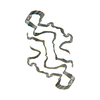
 Sample preparation
 Electron microscopy imaging
 FIELD EMISSION GUN / Accelerating voltage: 200 kV / Illumination mode: FLOOD BEAM
 Processing