 Movie
Movie Controller


+ Open data

- Basic information
| Entry | Database: PDB / ID: 7zdi | ||||||
|---|---|---|---|---|---|---|---|
| Title | PucB-LH2 complex from Rps. palustris | ||||||
 Components Components |
| ||||||
 Keywords Keywords | PHOTOSYNTHESIS / light harvesting complex 2 / LH2 / Rps. palustris / purple bacteria / cryo-EM / single particle analysis | ||||||
| Function / homology |  Function and homology information Function and homology informationorganelle inner membrane / plasma membrane light-harvesting complex / bacteriochlorophyll binding / photosynthesis, light reaction / metal ion binding / plasma membrane Similarity search - Function | ||||||
| Biological species |  Rhodopseudomonas palustris (phototrophic) Rhodopseudomonas palustris (phototrophic) | ||||||
| Method | ELECTRON MICROSCOPY / single particle reconstruction / cryo EM / Resolution: 2.9 Å | ||||||
 Authors Authors | Qian, P. / Cogdell, R.J. / Nguyen-Phan, T.C. | ||||||
| Funding support |  United Kingdom, 1items United Kingdom, 1items
| ||||||
 Citation Citation | Journal: Proc Natl Acad Sci U S A / Year: 2022 Title: Cryo-EM structures of light-harvesting 2 complexes from reveal the molecular origin of absorption tuning. Authors: Pu Qian / Cam T Nguyen-Phan / Alastair T Gardiner / Tristan I Croll / Aleksander W Roszak / June Southall / Philip J Jackson / Cvetelin Vasilev / Pablo Castro-Hartmann / Kasim Sader / C Neil ...Authors: Pu Qian / Cam T Nguyen-Phan / Alastair T Gardiner / Tristan I Croll / Aleksander W Roszak / June Southall / Philip J Jackson / Cvetelin Vasilev / Pablo Castro-Hartmann / Kasim Sader / C Neil Hunter / Richard J Cogdell /  Abstract: The genomes of some purple photosynthetic bacteria contain a multigene family encoding a series of α- and β-polypeptides that together form a heterogeneous antenna of light-harvesting 2 (LH2) ...The genomes of some purple photosynthetic bacteria contain a multigene family encoding a series of α- and β-polypeptides that together form a heterogeneous antenna of light-harvesting 2 (LH2) complexes. To unravel this complexity, we generated four sets of deletion mutants in , each encoding a single type of gene pair and enabling the purification of complexes designated as PucA-LH2, PucB-LH2, PucD-LH2, and PucE-LH2. The structures of all four purified LH2 complexes were determined by cryogenic electron microscopy (cryo-EM) at resolutions ranging from 2.7 to 3.6 Å. Uniquely, each of these complexes contains a hitherto unknown polypeptide, γ, that forms an extended undulating ribbon that lies in the plane of the membrane and that encloses six of the nine LH2 αβ-subunits. The γ-subunit, which is located near to the cytoplasmic side of the complex, breaks the C9 symmetry of the LH2 complex and binds six extra bacteriochlorophylls (BChls) that enhance the 800-nm absorption of each complex. The structures show that all four complexes have two complete rings of BChls, conferring absorption bands centered at 800 and 850 nm on the PucA-LH2, PucB-LH2, and PucE-LH2 complexes, but, unusually, the PucD-LH2 antenna has only a single strong near-infared (NIR) absorption peak at 803 nm. Comparison of the cryo-EM structures of these LH2 complexes reveals altered patterns of hydrogen bonds between LH2 αβ-side chains and the bacteriochlorin rings, further emphasizing the major role that H bonds play in spectral tuning of bacterial antenna complexes. #1: Journal: Acta Crystallogr., Sect. D: Biol. Crystallogr. / Year: 2018Title: Real-space refinement in PHENIX for cryo-EM and crystallography Authors: Qian, P. / Cogdell, R.J. #2: Journal: To Be PublishedTitle: Cryo-EM structures of light harvesting complex 2 from Rhodopseudomonas palustris: a molecular origin of spectroscopic variation Authors: Qian, P. / Cogdell, R.J. | ||||||
| History |
|
- Structure visualization
| Structure viewer | Molecule: MolmilJmol/JSmol |
|---|
- Downloads & links
-Download
| PDBx/mmCIF format | 7zdi.cif.gz | 320.2 KB | Display | PDBx/mmCIF format |
|---|---|---|---|---|
| PDB format | pdb7zdi.ent.gz | 224.3 KB | Display | PDB format |
| PDBx/mmJSON format | 7zdi.json.gz | Tree view | PDBx/mmJSON format | |
| Others |  Other downloads Other downloads |
-Validation report
| Arichive directory | https://data.pdbj.org/pub/pdb/validation_reports/zd/7zdiftp://data.pdbj.org/pub/pdb/validation_reports/zd/7zdi | HTTPS FTP |
|---|
-Related structure data
| Related structure data |  14650MC  7zcuC 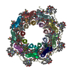 7ze3C  7ze8C M: map data used to model this data C: citing same article ( |
|---|---|
| Similar structure data |


-Links
 PDBj
PDBj- Assembly
| Deposited unit | 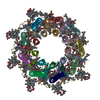
|
|---|---|
| 1 |
|
-Components
| #1: Protein | Mass: 6873.004 Da / Num. of mol.: 9 / Source method: isolated from a natural source / Source: (natural) Rhodopseudomonas palustris (phototrophic) / References: UniProt: A0A323UGK7#2: Protein | Mass: 5738.550 Da / Num. of mol.: 9 / Source method: isolated from a natural source / Source: (natural) Rhodopseudomonas palustris (phototrophic) / References: UniProt: A0A2R4GZC1#3: Protein | | Mass: 10848.179 Da / Num. of mol.: 1 / Source method: isolated from a natural source / Source: (natural) Rhodopseudomonas palustris (phototrophic) / References: UniProt: A0A2R4GRK2#4: Chemical | ChemComp-BCL /   Mass: 911.504 Da / Num. of mol.: 33 / Source method: obtained synthetically / Formula: C55H74MgN4O6 / Feature type: SUBJECT OF INVESTIGATION Mass: 911.504 Da / Num. of mol.: 33 / Source method: obtained synthetically / Formula: C55H74MgN4O6 / Feature type: SUBJECT OF INVESTIGATION#5: Chemical | ChemComp-IRM /   Mass: 554.888 Da / Num. of mol.: 9 / Source method: obtained synthetically / Formula: C40H58O / Feature type: SUBJECT OF INVESTIGATION Mass: 554.888 Da / Num. of mol.: 9 / Source method: obtained synthetically / Formula: C40H58O / Feature type: SUBJECT OF INVESTIGATIONHas ligand of interest | Y | Has protein modification | Y | |
|---|
-Experimental details
-Experiment
| Experiment | Method: ELECTRON MICROSCOPY |
|---|---|
| EM experiment | Aggregation state: PARTICLE / 3D reconstruction method: single particle reconstruction |
- Sample preparation
| Component | Name: PucB-LH2 from Rps. palustris / Type: COMPLEX / Details: Delt-pucBA(aced) / Entity ID: #1-#3 / Source: NATURAL |
|---|---|
| Source (natural) | Organism: Rhodopseudomonas palustris ATCC 17001 (phototrophic) |
| Buffer solution | pH: 8 / Details: 0.1% LDAO in 20 mM Tris.Cl pH 8.0 |
| Buffer component | Conc.: 20 mM / Name: Tris.HCl / Formula: C4H11NO3 |
| Specimen | Conc.: 7 mg/ml / Embedding applied: NO / Shadowing applied: NO / Staining applied: NO / Vitrification applied: YES / Details: protein was purified using LDAO detergent. |
| Specimen support | Grid material: COPPER / Grid mesh size: 300 divisions/in. / Grid type: Quantifoil R1.2/1.3 |
| Vitrification | Instrument: FEI VITROBOT MARK IV / Cryogen name: ETHANE / Humidity: 100 % / Chamber temperature: 277 K Details: Chamber humidity: 100%; Chamber temperature: 4 oC; Blotting time: 2.5; Blotting force: 3; Wait time: 30 sec |
- Electron microscopy imaging
| Experimental equipment |  Model: Titan Krios / Image courtesy: FEI Company |
|---|---|
| Microscopy | Model: FEI TITAN KRIOS |
| Electron gun | Electron source:  FIELD EMISSION GUN / Accelerating voltage: 300 kV / Illumination mode: FLOOD BEAM FIELD EMISSION GUN / Accelerating voltage: 300 kV / Illumination mode: FLOOD BEAM |
| Electron lens | Mode: BRIGHT FIELD / Nominal magnification: 120000 X / Nominal defocus max: 2400 nm / Nominal defocus min: 800 nm / Cs: 2.7 mm / C2 aperture diameter: 50 µm / Alignment procedure: COMA FREE |
| Specimen holder | Cryogen: NITROGEN / Specimen holder model: FEI TITAN KRIOS AUTOGRID HOLDER / Temperature (min): 80 K |
| Image recording | Average exposure time: 12.21 sec. / Electron dose: 44.1 e/Å2 / Film or detector model: FEI FALCON IV (4k x 4k) / Num. of grids imaged: 1 / Num. of real images: 5593 / Details: Images were collected in AFIS mode. |
| EM imaging optics | Details: No energy filter was applied. |
- Processing
| Software |
| ||||||||||||||||||||||||||||||||||||
|---|---|---|---|---|---|---|---|---|---|---|---|---|---|---|---|---|---|---|---|---|---|---|---|---|---|---|---|---|---|---|---|---|---|---|---|---|---|
| EM software |
| ||||||||||||||||||||||||||||||||||||
| CTF correction | Type: NONE | ||||||||||||||||||||||||||||||||||||
| Particle selection | Num. of particles selected: 1946434 | ||||||||||||||||||||||||||||||||||||
| Symmetry | Point symmetry: C1 (asymmetric) | ||||||||||||||||||||||||||||||||||||
| 3D reconstruction | Resolution: 2.9 Å / Resolution method: FSC 0.143 CUT-OFF / Num. of particles: 402668 / Algorithm: BACK PROJECTION / Num. of class averages: 1 / Symmetry type: POINT | ||||||||||||||||||||||||||||||||||||
| Atomic model building | Protocol: FLEXIBLE FIT / Space: REAL | ||||||||||||||||||||||||||||||||||||
| Refinement | Cross valid method: NONE Stereochemistry target values: GeoStd + Monomer Library + CDL v1.2 | ||||||||||||||||||||||||||||||||||||
| Displacement parameters | Biso mean: 39.44 Å2 | ||||||||||||||||||||||||||||||||||||
| Refine LS restraints |
|
