Movie
Movie Controller

[English] 日本語
 Yorodumi
- PDB-7zcw: Cryo-EM structure of GMPCPP-microtubules in complex with VASH2-SVBP -
+ Open data

- Basic information
| Entry | Database: PDB / ID: 7zcw | ||||||
|---|---|---|---|---|---|---|---|
| Title | Cryo-EM structure of GMPCPP-microtubules in complex with VASH2-SVBP | ||||||
 Components Components |
| ||||||
 Keywords Keywords | PROTEIN BINDING / Microtubule / Enzyme / Complex / Detyrosination | ||||||
| Function / homology |  Function and homology information Function and homology informationcell-cell fusion / regulation of metallopeptidase activity / tubulinyl-Tyr carboxypeptidase / tubulin-tyrosine carboxypeptidase activity / syncytium formation by cell-cell fusion / Post-chaperonin tubulin folding pathway / Cilium Assembly / cytoskeleton-dependent intracellular transport / Carboxyterminal post-translational modifications of tubulin / Microtubule-dependent trafficking of connexons from Golgi to the plasma membrane ...cell-cell fusion / regulation of metallopeptidase activity / tubulinyl-Tyr carboxypeptidase / tubulin-tyrosine carboxypeptidase activity / syncytium formation by cell-cell fusion / Post-chaperonin tubulin folding pathway / Cilium Assembly / cytoskeleton-dependent intracellular transport / Carboxyterminal post-translational modifications of tubulin / Microtubule-dependent trafficking of connexons from Golgi to the plasma membrane / Sealing of the nuclear envelope (NE) by ESCRT-III / Intraflagellar transport / Formation of tubulin folding intermediates by CCT/TriC / embryonic brain development / Gap junction assembly / Kinesins / positive regulation of axon guidance / Prefoldin mediated transfer of substrate to CCT/TriC / negative regulation of endothelial cell migration / Assembly and cell surface presentation of NMDA receptors / COPI-independent Golgi-to-ER retrograde traffic / labyrinthine layer blood vessel development / peptidase activator activity / COPI-dependent Golgi-to-ER retrograde traffic / axon development / Recycling pathway of L1 / RHOH GTPase cycle / protein secretion / regulation of angiogenesis / microtubule-based process / RHO GTPases activate IQGAPs / Hedgehog 'off' state / intercellular bridge / COPI-mediated anterograde transport / cytoplasmic microtubule / Activation of AMPK downstream of NMDARs / metallocarboxypeptidase activity / positive regulation of endothelial cell proliferation / MHC class II antigen presentation / negative regulation of protein ubiquitination / Recruitment of NuMA to mitotic centrosomes / Mitotic Prometaphase / HSP90 chaperone cycle for steroid hormone receptors (SHR) in the presence of ligand / cellular response to interleukin-4 / EML4 and NUDC in mitotic spindle formation / Resolution of Sister Chromatid Cohesion / Translocation of SLC2A4 (GLUT4) to the plasma membrane / RHO GTPases Activate Formins / cerebral cortex development / PKR-mediated signaling / structural constituent of cytoskeleton / modulation of chemical synaptic transmission / microtubule cytoskeleton organization / Schaffer collateral - CA1 synapse / neuron migration / positive regulation of angiogenesis / HCMV Early Events / apical part of cell / Aggrephagy / The role of GTSE1 in G2/M progression after G2 checkpoint / mitotic spindle / Separation of Sister Chromatids / mitotic cell cycle / double-stranded RNA binding / microtubule cytoskeleton / actin binding / microtubule binding / cytoskeleton / microtubule / Hydrolases; Acting on acid anhydrides; Acting on GTP to facilitate cellular and subcellular movement / cilium / protein heterodimerization activity / cell division / GTPase activity / ubiquitin protein ligase binding / GTP binding / structural molecule activity / proteolysis / extracellular region / metal ion binding / nucleus / cytoplasm / cytosol Similarity search - Function | ||||||
| Biological species |  Homo sapiens (human) Homo sapiens (human) | ||||||
| Method | ELECTRON MICROSCOPY / single particle reconstruction / cryo EM / Resolution: 3.6 Å | ||||||
 Authors Authors | Choi, S.R. / Blum, T. / Steinmetz, M.O. | ||||||
| Funding support |  Switzerland, 1items Switzerland, 1items
| ||||||
 Citation Citation | Journal: J Cell Biol / Year: 2023 Title: VASH1-SVBP and VASH2-SVBP generate different detyrosination profiles on microtubules. Authors: Sacnicte Ramirez-Rios / Sung Ryul Choi / Chadni Sanyal / Thorsten B Blum / Christophe Bosc / Fatma Krichen / Eric Denarier / Jean-Marc Soleilhac / Béatrice Blot / Carsten Janke / Virginie ...Authors: Sacnicte Ramirez-Rios / Sung Ryul Choi / Chadni Sanyal / Thorsten B Blum / Christophe Bosc / Fatma Krichen / Eric Denarier / Jean-Marc Soleilhac / Béatrice Blot / Carsten Janke / Virginie Stoppin-Mellet / Maria M Magiera / Isabelle Arnal / Michel O Steinmetz / Marie-Jo Moutin /  Abstract: The detyrosination/tyrosination cycle of α-tubulin is critical for proper cell functioning. VASH1-SVBP and VASH2-SVBP are ubiquitous enzymes involved in microtubule detyrosination, whose mode of ...The detyrosination/tyrosination cycle of α-tubulin is critical for proper cell functioning. VASH1-SVBP and VASH2-SVBP are ubiquitous enzymes involved in microtubule detyrosination, whose mode of action is little known. Here, we show in reconstituted systems and cells that VASH1-SVBP and VASH2-SVBP drive the global and local detyrosination of microtubules, respectively. We solved the cryo-electron microscopy structure of VASH2-SVBP bound to microtubules, revealing a different microtubule-binding configuration of its central catalytic region compared to VASH1-SVBP. We show that the divergent mode of detyrosination between the two enzymes is correlated with the microtubule-binding properties of their disordered N- and C-terminal regions. Specifically, the N-terminal region is responsible for a significantly longer residence time of VASH2-SVBP on microtubules compared to VASH1-SVBP. We suggest that this VASH region is critical for microtubule detachment and diffusion of VASH-SVBP enzymes on lattices. Our results suggest a mechanism by which VASH1-SVBP and VASH2-SVBP could generate distinct microtubule subpopulations and confined areas of detyrosinated lattices to drive various microtubule-based cellular functions. | ||||||
| History |
|
- Structure visualization
| Structure viewer | Molecule: MolmilJmol/JSmol |
|---|
- Downloads & links
-Download
| PDBx/mmCIF format | 7zcw.cif.gz | 937.1 KB | Display | PDBx/mmCIF format |
|---|---|---|---|---|
| PDB format | pdb7zcw.ent.gz | 779.3 KB | Display | PDB format |
| PDBx/mmJSON format | 7zcw.json.gz | Tree view | PDBx/mmJSON format | |
| Others |  Other downloads Other downloads |
-Validation report
| Arichive directory | https://data.pdbj.org/pub/pdb/validation_reports/zc/7zcwftp://data.pdbj.org/pub/pdb/validation_reports/zc/7zcw | HTTPS FTP |
|---|
-Related structure data
| Related structure data | 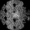 14634MC M: map data used to model this data C: citing same article ( |
|---|---|
| Similar structure data |
-Links
 PDBj
PDBj


- Assembly
| Deposited unit | 
|
|---|---|
| 1 |
|
-Components
-Protein , 4 types, 8 molecules AEBFGHCD
| #1: Protein | Mass: 50204.445 Da / Num. of mol.: 2 / Source method: isolated from a natural source / Source: (natural) Homo sapiens (human) / Cell line: Hela / References: UniProt: P68363#2: Protein | Mass: 49907.770 Da / Num. of mol.: 4 / Source method: isolated from a natural source / Source: (natural) Homo sapiens (human) / Cell line: HELA / References: UniProt: Q9BVA1#3: Protein | | Mass: 40488.914 Da / Num. of mol.: 1 / Mutation: C158A Source method: isolated from a genetically manipulated source Source: (gene. exp.) Homo sapiens (human) / Gene: VASH2, VASHL / Production host:  #4: Protein | | Mass: 8780.000 Da / Num. of mol.: 1 Source method: isolated from a genetically manipulated source Details: Recombinant C-terminal his6-tag / Source: (gene. exp.) Homo sapiens (human) / Gene: SVBP, CCDC23 / Production host: |
|---|
-Non-polymers , 3 types, 12 molecules 


| #5: Chemical |  Mass: 523.180 Da / Num. of mol.: 2 / Source method: obtained synthetically / Formula: C10H16N5O14P3 / Comment: GTP, energy-carrying molecule*YM Mass: 523.180 Da / Num. of mol.: 2 / Source method: obtained synthetically / Formula: C10H16N5O14P3 / Comment: GTP, energy-carrying molecule*YM#6: Chemical | ChemComp-MG /  Mass: 24.305 Da / Num. of mol.: 6 / Source method: obtained synthetically / Formula: Mg Mass: 24.305 Da / Num. of mol.: 6 / Source method: obtained synthetically / Formula: Mg#7: Chemical | ChemComp-G2P /  Mass: 521.208 Da / Num. of mol.: 4 / Source method: obtained synthetically / Formula: C11H18N5O13P3 / Comment: GMP-CPP, energy-carrying molecule analogue*YM Mass: 521.208 Da / Num. of mol.: 4 / Source method: obtained synthetically / Formula: C11H18N5O13P3 / Comment: GMP-CPP, energy-carrying molecule analogue*YM |
|---|
-Details
| Has ligand of interest | N |
|---|
-Experimental details
-Experiment
| Experiment | Method: ELECTRON MICROSCOPY |
|---|---|
| EM experiment | Aggregation state: HELICAL ARRAY / 3D reconstruction method: single particle reconstruction |
- Sample preparation
| Component | Name: VASH2-SVBP complex bound to the microtubule / Type: COMPLEX / Entity ID: #1-#4 / Source: RECOMBINANT | ||||||||||||||||||||
|---|---|---|---|---|---|---|---|---|---|---|---|---|---|---|---|---|---|---|---|---|---|
| Molecular weight | Value: 0.38 MDa / Experimental value: NO | ||||||||||||||||||||
| Source (natural) | Organism: Homo sapiens (human) | ||||||||||||||||||||
| Source (recombinant) | Organism: | ||||||||||||||||||||
| Buffer solution | pH: 7.5 | ||||||||||||||||||||
| Buffer component |
| ||||||||||||||||||||
| Specimen | Conc.: 2 mg/ml / Embedding applied: NO / Shadowing applied: NO / Staining applied: NO / Vitrification applied: YES / Details: Mixture of VASH2-SVBP and microtubules | ||||||||||||||||||||
| Specimen support | Grid material: COPPER / Grid mesh size: 300 divisions/in. / Grid type: Quantifoil R2/1 | ||||||||||||||||||||
| Vitrification | Instrument: FEI VITROBOT MARK III / Cryogen name: ETHANE / Humidity: 100 % / Chamber temperature: 298 K |
- Electron microscopy imaging
| Experimental equipment |  Model: Titan Krios / Image courtesy: FEI Company |
|---|---|
| Microscopy | Model: FEI TITAN KRIOS |
| Electron gun | Electron source:  FIELD EMISSION GUN / Accelerating voltage: 300 kV / Illumination mode: FLOOD BEAM FIELD EMISSION GUN / Accelerating voltage: 300 kV / Illumination mode: FLOOD BEAM |
| Electron lens | Mode: BRIGHT FIELD / Nominal defocus max: 2200 nm / Nominal defocus min: 600 nm |
| Image recording | Average exposure time: 0.2 sec. / Electron dose: 1.4 e/Å2 / Detector mode: COUNTING / Film or detector model: GATAN K2 SUMMIT (4k x 4k) / Num. of grids imaged: 1 / Num. of real images: 3345 |
- Processing
| EM software |
| ||||||||||||||||||||||||||||||||||||||||||||
|---|---|---|---|---|---|---|---|---|---|---|---|---|---|---|---|---|---|---|---|---|---|---|---|---|---|---|---|---|---|---|---|---|---|---|---|---|---|---|---|---|---|---|---|---|---|
| CTF correction | Type: PHASE FLIPPING AND AMPLITUDE CORRECTION | ||||||||||||||||||||||||||||||||||||||||||||
| Particle selection | Num. of particles selected: 153041 | ||||||||||||||||||||||||||||||||||||||||||||
| 3D reconstruction | Resolution: 3.6 Å / Resolution method: FSC 0.143 CUT-OFF / Num. of particles: 96922 / Num. of class averages: 1 / Symmetry type: POINT | ||||||||||||||||||||||||||||||||||||||||||||
| Atomic model building | B value: 54.76 / Protocol: RIGID BODY FIT | ||||||||||||||||||||||||||||||||||||||||||||
| Atomic model building | 3D fitting-ID: 1 / Source name: PDB / Type: experimental model
|