 Movie
Movie Controller


+ Open data

- Basic information
| Entry | Database: PDB / ID: 7zct | ||||||
|---|---|---|---|---|---|---|---|
| Title | Structure of the red fluorescent protein mScarlet3 at pH 7.5 | ||||||
 Components Components | Red fluorescent protein drFP583 | ||||||
 Keywords Keywords | FLUORESCENT PROTEIN / Red fluorescent protein / high fluorescence quantum yield / efficient chromophore maturation | ||||||
| Function / homology | Green fluorescent protein, GFP / Green fluorescent protein-related / Green fluorescent protein / Green fluorescent protein / bioluminescence / generation of precursor metabolites and energy / Red fluorescent protein drFP583 Function and homology information Function and homology information | ||||||
| Biological species |  Discosoma sp. (sea anemone) Discosoma sp. (sea anemone) | ||||||
| Method |  X-RAY DIFFRACTION / SYNCHROTRON / MOLECULAR REPLACEMENT / Resolution: 1.33 Å X-RAY DIFFRACTION / SYNCHROTRON / MOLECULAR REPLACEMENT / Resolution: 1.33 Å | ||||||
 Authors Authors | Aumonier, S. / Dupuy, J. / Royant, A. | ||||||
| Funding support |  France, 1items France, 1items
| ||||||
 Citation Citation | Journal: Nat.Methods / Year: 2023 Title: mScarlet3: a brilliant and fast-maturing red fluorescent protein. Authors: Gadella Jr., T.W.J. / van Weeren, L. / Stouthamer, J. / Hink, M.A. / Wolters, A.H.G. / Giepmans, B.N.G. / Aumonier, S. / Dupuy, J. / Royant, A. #1: Journal: Nat Methods / Year: 2017Title: mScarlet: a bright monomeric red fluorescent protein for cellular imaging. Authors: Bindels, D.S. / Haarbosch, L. / van Weeren, L. / Postma, M. / Wiese, K.E. / Mastop, M. / Aumonier, S. / Gotthard, G. / Royant, A. / Hink, M.A. / Gadella, T.W. | ||||||
| History |
|
- Structure visualization
| Structure viewer | Molecule: MolmilJmol/JSmol |
|---|
- Downloads & links
-Download
| PDBx/mmCIF format | 7zct.cif.gz | 109.1 KB | Display | PDBx/mmCIF format |
|---|---|---|---|---|
| PDB format | pdb7zct.ent.gz | 82.5 KB | Display | PDB format |
| PDBx/mmJSON format | 7zct.json.gz | Tree view | PDBx/mmJSON format | |
| Others |  Other downloads Other downloads |
-Validation report
| Arichive directory | https://data.pdbj.org/pub/pdb/validation_reports/zc/7zctftp://data.pdbj.org/pub/pdb/validation_reports/zc/7zct | HTTPS FTP |
|---|
-Related structure data
| Related structure data |  5lk4S S: Starting model for refinement |
|---|---|
| Similar structure data |
-Links
 PDBj
PDBj

- Assembly
| Deposited unit | 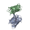
| ||||||||
|---|---|---|---|---|---|---|---|---|---|
| 1 | 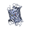
| ||||||||
| 2 | 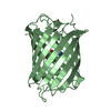
| ||||||||
| Unit cell |
|
-Components
| #1: Protein | Mass: 25865.129 Da / Num. of mol.: 2 Source method: isolated from a genetically manipulated source Details: Residue NRQ67 is formed by the autocatalytic cyclisation of the three consecutive amino acid residues M67, Y68 and G69 Source: (gene. exp.) Discosoma sp. (sea anemone) / Production host:  #2: Water | ChemComp-HOH / |  Mass: 18.015 Da / Num. of mol.: 244 / Source method: isolated from a natural source / Formula: H2O Mass: 18.015 Da / Num. of mol.: 244 / Source method: isolated from a natural source / Formula: H2OHas ligand of interest | Y | |
|---|
-Experimental details
-Experiment
| Experiment | Method: X-RAY DIFFRACTION / Number of used crystals: 1 |
|---|
- Sample preparation
| Crystal | Density Matthews: 1.77 Å3/Da / Density % sol: 30.42 % |
|---|---|
| Crystal grow | Temperature: 293 K / Method: vapor diffusion, hanging drop / pH: 7.5 Details: 0.1 M sodium chloride, 0.1 M, 0.1 M HEPES pH 7.5, 1.6 M Ammonium sulfate |
-Data collection
| Diffraction | Mean temperature: 100 K / Serial crystal experiment: N |
|---|---|
| Diffraction source | Source: SYNCHROTRON / Site: ESRF / Beamline: ID30B / Wavelength: 0.9686 Å |
| Detector | Type: DECTRIS PILATUS3 6M / Detector: PIXEL / Date: Sep 10, 2020 |
| Radiation | Monochromator: CHANNEL-CUT SI(111) CRYSTAL / Protocol: SINGLE WAVELENGTH / Monochromatic (M) / Laue (L): M / Scattering type: x-ray |
| Radiation wavelength | Wavelength: 0.9686 Å / Relative weight: 1 |
| Reflection | Resolution: 1.33→42.78 Å / Num. obs: 91873 / % possible obs: 97.5 % / Redundancy: 3.8 % / Biso Wilson estimate: 16.7 Å2 / CC1/2: 0.994 / Rsym value: 0.097 / Net I/σ(I): 6 |
| Reflection shell | Resolution: 1.33→1.38 Å / Mean I/σ(I) obs: 0.7 / Num. unique obs: 9289 / CC1/2: 0.548 / Rsym value: 0.929 / % possible all: 99.1 |
- Processing
| Software |
| ||||||||||||||||||||||||||||||||||||||||||||||||||||||||||||
|---|---|---|---|---|---|---|---|---|---|---|---|---|---|---|---|---|---|---|---|---|---|---|---|---|---|---|---|---|---|---|---|---|---|---|---|---|---|---|---|---|---|---|---|---|---|---|---|---|---|---|---|---|---|---|---|---|---|---|---|---|---|
| Refinement | Method to determine structure: MOLECULAR REPLACEMENT Starting model: 5LK4 Resolution: 1.33→42.78 Å / Cor.coef. Fo:Fc: 0.969 / Cor.coef. Fo:Fc free: 0.955 / SU B: 1.84 / SU ML: 0.07 / Cross valid method: THROUGHOUT / σ(F): 0 / ESU R: 0.061 / ESU R Free: 0.063 / Stereochemistry target values: MAXIMUM LIKELIHOOD Details: HYDROGENS HAVE BEEN ADDED IN THE RIDING POSITIONS U VALUES : REFINED INDIVIDUALLY
| ||||||||||||||||||||||||||||||||||||||||||||||||||||||||||||
| Solvent computation | Ion probe radii: 0.8 Å / Shrinkage radii: 0.8 Å / VDW probe radii: 1.2 Å / Solvent model: MASK | ||||||||||||||||||||||||||||||||||||||||||||||||||||||||||||
| Displacement parameters | Biso max: 67.36 Å2 / Biso mean: 18.863 Å2 / Biso min: 12.02 Å2
| ||||||||||||||||||||||||||||||||||||||||||||||||||||||||||||
| Refinement step | Cycle: final / Resolution: 1.33→42.78 Å
| ||||||||||||||||||||||||||||||||||||||||||||||||||||||||||||
| Refine LS restraints |
| ||||||||||||||||||||||||||||||||||||||||||||||||||||||||||||
| LS refinement shell | Resolution: 1.33→1.362 Å / Rfactor Rfree error: 0
|
