[English] 日本語
Yorodumi
- PDB-7zai: Cryo-EM structure of a Pyrococcus abyssi 30S bound to Met-initiat... -

+
Open data


ID or keywords:

Loading...

-
Basic information

Entry
Database: PDB / ID: 7zai
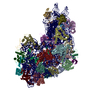
TitleCryo-EM structure of a Pyrococcus abyssi 30S bound to Met-initiator tRNA, mRNA and aIF1A.
Components
  • (30S ribosomal protein ...) x 25
  • (50S ribosomal protein ...) x 2
  • 16S rRNA
  • Translation initiation factor 1A
  • Zn-ribbon RNA-binding protein involved in translation
  • mRNA
  • tRNA-MET
KeywordsTRANSLATION / Initiation complex / translation initiation / small ribosomal subunit / aIF5b
Function / homology
Function and homology information


ribonuclease P activity / tRNA 5'-leader removal / translation initiation factor activity / ribosome biogenesis / small ribosomal subunit / tRNA binding / rRNA binding / ribosome / structural constituent of ribosome / translation ...ribonuclease P activity / tRNA 5'-leader removal / translation initiation factor activity / ribosome biogenesis / small ribosomal subunit / tRNA binding / rRNA binding / ribosome / structural constituent of ribosome / translation / ribonucleoprotein complex / RNA binding / zinc ion binding / cytosol / cytoplasm
Similarity search - Function
Small zinc finger protein HVO_2753-like, zinc-binding pocket / Small zinc finger protein HVO_2753-like / Small zinc finger protein HVO_2753-like, Zn-binding pocket / Ribosomal protein S27ae / Ribosomal protein S9, archaeal / Ribosomal protein S13, archaeal / Ribosomal protein S17, archaeal / Ribosomal protein S6e, archaeal / Ribosomal protein S4, archaeal / Ribosomal protein S12, archaea ...Small zinc finger protein HVO_2753-like, zinc-binding pocket / Small zinc finger protein HVO_2753-like / Small zinc finger protein HVO_2753-like, Zn-binding pocket / Ribosomal protein S27ae / Ribosomal protein S9, archaeal / Ribosomal protein S13, archaeal / Ribosomal protein S17, archaeal / Ribosomal protein S6e, archaeal / Ribosomal protein S4, archaeal / Ribosomal protein S12, archaea / Ribosomal protein S3, archaeal / 30S ribosomal protein S3Ae / : / Ribosomal protein S7, archaeal / Ribosomal protein S19e, archaeal / Ribosomal protein S11, archaeal / Ribosomal protein S2, archaeal / Ribosomal protein S14, type Z, archaeal / Ribosomal protein S8e, archaeal / Translation initiation factor 1A (eIF-1A), conserved site / Eukaryotic initiation factor 1A signature. / eukaryotic translation initiation factor 1A / Translation initiation factor 1A (eIF-1A) / RNA-binding domain, S1, IF1 type / Translation initiation factor 1A / IF-1 / S1 domain IF1 type profile. / Ribosomal protein L7Ae, archaea / Ribosomal protein S5, eukaryotic/archaeal / Ribosomal protein S19e, conserved site / Ribosomal protein S19e signature. / Ribosomal protein S10, eukaryotic/archaeal / Ribosomal protein S27a / Ribosomal protein S27a / Ribosomal protein S27a / Ribosomal protein S2, eukaryotic/archaeal / Ribosomal protein S17e, conserved site / Ribosomal protein S17e signature. / 40S ribosomal protein S29/30S ribosomal protein S14 type Z / Ribosomal protein S3, eukaryotic/archaeal / Ribosomal protein S3Ae, conserved site / Ribosomal protein S3Ae signature. / Ribosomal protein S19e / Ribosomal protein S19e / Ribosomal_S19e / Ribosomal protein S27e signature. / Ribosomal protein S4e, N-terminal, conserved site / Ribosomal protein S4e signature. / Ribosomal protein S8e, conserved site / Ribosomal protein S8e signature. / Ribosomal protein S19A/S15e / Ribosomal protein S17e / Ribosomal protein S17e-like superfamily / Ribosomal S17 / Ribosomal S24e conserved site / Ribosomal protein S24e signature. / Ribosomal protein S4e, N-terminal / RS4NT (NUC023) domain / Ribosomal protein S4, KOW domain / Ribosomal protein S4e / Ribosomal protein S4e, central region / Ribosomal protein S4e, central domain superfamily / Ribosomal family S4e / Ribosomal protein S6/S6e/A/B/2, conserved site / Ribosomal protein S6e signature. / Ribosomal protein S23, eukaryotic/archaeal / Ribosomal protein S24e / Ribosomal protein S24e / Ribosomal protein S27 / Ribosomal protein S27, zinc-binding domain superfamily / Ribosomal protein S27 / Ribosomal protein S8e / Ribosomal protein S17, archaeal/eukaryotic / Ribosomal protein S28e conserved site / Ribosomal protein S28e signature. / Ribosomal protein S3Ae / Ribosomal S3Ae family / Ribosomal S3Ae family / Ribosomal protein S28e / Ribosomal protein S28e / Ribosomal protein S6e / Ribosomal protein S5/S7, eukaryotic/archaeal / Ribosomal protein S6e / Ribosomal protein S6e / Ribosomal protein S13/S15, N-terminal / Ribosomal protein S15P / Ribosomal S13/S15 N-terminal domain / Ribosomal S13/S15 N-terminal domain / Ribosomal protein S4/S9, eukaryotic/archaeal / Ribosomal protein L7Ae conserved site / Ribosomal protein L7Ae signature. / Ribosomal protein S8e/ribosomal biogenesis NSA2 / Ribosomal protein S8e / Ribosomal protein L7Ae/L8/Nhp2 family / Ribosomal protein S14/S29 / Ribosomal protein L7Ae/L30e/S12e/Gadd45 / Ribosomal protein L7Ae/L30e/S12e/Gadd45 family / 50S ribosomal protein L30e-like / Ribosomal protein S2 signature 2. / K Homology domain / K homology RNA-binding domain
Similarity search - Domain/homology
METHIONINE / : / : / RNA / RNA (> 10) / RNA (> 100) / RNA (> 1000) / Zn-ribbon RNA-binding protein involved in translation / Small ribosomal subunit protein eS28 / Small ribosomal subunit protein eS31 ...METHIONINE / : / : / RNA / RNA (> 10) / RNA (> 100) / RNA (> 1000) / Zn-ribbon RNA-binding protein involved in translation / Small ribosomal subunit protein eS28 / Small ribosomal subunit protein eS31 / Small ribosomal subunit protein uS4 / Small ribosomal subunit protein uS12 / Large ribosomal subunit protein eL8 / Small ribosomal subunit protein uS11 / Small ribosomal subunit protein uS14 / Small ribosomal subunit protein eS32 / Small ribosomal subunit protein eS27 / Small ribosomal subunit protein eS24 / Small ribosomal subunit protein eS6 / Small ribosomal subunit protein eS8 / Small ribosomal subunit protein eS17 / Small ribosomal subunit protein eS19 / Small ribosomal subunit protein uS10 / Small ribosomal subunit protein uS7 / Translation initiation factor 1A / Small ribosomal subunit protein uS2 / Small ribosomal subunit protein uS9 / Small ribosomal subunit protein uS13 / Small ribosomal subunit protein uS19 / Small ribosomal subunit protein uS3 / Small ribosomal subunit protein uS17 / Small ribosomal subunit protein eS4 / Small ribosomal subunit protein uS8 / Small ribosomal subunit protein uS5 / Small ribosomal subunit protein eS1 / Small ribosomal subunit protein uS15
Similarity search - Component
Biological speciesPyrococcus abyssi GE5 (archaea)
Pyrococcus abyssi (archaea)
MethodELECTRON MICROSCOPY / single particle reconstruction / cryo EM / Resolution: 2.6 Å
AuthorsCoureux, P.D. / Bourgeois, G. / Mechulam, Y. / Schmitt, E. / Kazan, R.
Funding support France, 1items
OrganizationGrant numberCountry
Agence Nationale de la Recherche (ANR) France
CitationJournal: Nucleic Acids Res / Year: 2022
Title: Role of aIF5B in archaeal translation initiation.
Authors: Ramy Kazan / Gabrielle Bourgeois / Christine Lazennec-Schurdevin / Eric Larquet / Yves Mechulam / Pierre-Damien Coureux / Emmanuelle Schmitt /
Abstract: In eukaryotes and in archaea late steps of translation initiation involve the two initiation factors e/aIF5B and e/aIF1A. In eukaryotes, the role of eIF5B in ribosomal subunit joining is established ...In eukaryotes and in archaea late steps of translation initiation involve the two initiation factors e/aIF5B and e/aIF1A. In eukaryotes, the role of eIF5B in ribosomal subunit joining is established and structural data showing eIF5B bound to the full ribosome were obtained. To achieve its function, eIF5B collaborates with eIF1A. However, structural data illustrating how these two factors interact on the small ribosomal subunit have long been awaited. The role of the archaeal counterparts, aIF5B and aIF1A, remains to be extensively addressed. Here, we study the late steps of Pyrococcus abyssi translation initiation. Using in vitro reconstituted initiation complexes and light scattering, we show that aIF5B bound to GTP accelerates subunit joining without the need for GTP hydrolysis. We report the crystallographic structures of aIF5B bound to GDP and GTP and analyze domain movements associated to these two nucleotide states. Finally, we present the cryo-EM structure of an initiation complex containing 30S bound to mRNA, Met-tRNAiMet, aIF5B and aIF1A at 2.7 Å resolution. Structural data shows how archaeal 5B and 1A factors cooperate to induce a conformation of the initiator tRNA favorable to subunit joining. Archaeal and eukaryotic features of late steps of translation initiation are discussed.
History
DepositionMar 22, 2022Deposition site: PDBE / Processing site: PDBE
Revision 1.0Jul 13, 2022Provider: repository / Type: Initial release
Revision 1.1Aug 31, 2022Group: Database references / Category: citation / citation_author
Item: _citation.journal_volume / _citation.page_first ..._citation.journal_volume / _citation.page_first / _citation.page_last / _citation_author.identifier_ORCID
Revision 1.2Apr 24, 2024Group: Data collection / Refinement description
Category: chem_comp_atom / chem_comp_bond ...chem_comp_atom / chem_comp_bond / em_3d_fitting_list / pdbx_initial_refinement_model
Item: _em_3d_fitting_list.accession_code / _em_3d_fitting_list.initial_refinement_model_id ..._em_3d_fitting_list.accession_code / _em_3d_fitting_list.initial_refinement_model_id / _em_3d_fitting_list.source_name / _em_3d_fitting_list.type
Revision 1.3Nov 13, 2024Group: Data collection / Structure summary / Category: em_admin / pdbx_entry_details
Item: _em_admin.last_update / _pdbx_entry_details.has_protein_modification

-
Structure visualization

Structure viewerMolecule:
MolmilJmol/JSmol

Downloads & links

-
Assembly

Deposited unit
2: 16S rRNA
A: 30S ribosomal protein S3Ae
B: 30S ribosomal protein S2
C: Zn-ribbon RNA-binding protein involved in translation
D: 30S ribosomal protein S4
E: 30S ribosomal protein S4e
F: 30S ribosomal protein S5
G: 30S ribosomal protein S6e
H: 30S ribosomal protein S7
I: 30S ribosomal protein S8
J: 30S ribosomal protein S8e
K: 30S ribosomal protein S9
L: 30S ribosomal protein S10
M: 30S ribosomal protein S11
N: 30S ribosomal protein S12
O: 30S ribosomal protein S13
P: 30S ribosomal protein S14 type Z
Q: 30S ribosomal protein S15
R: 30S ribosomal protein S17
S: 30S ribosomal protein S17e
T: 30S ribosomal protein S19
U: 30S ribosomal protein S19e
V: 30S ribosomal protein S24e
W: 30S ribosomal protein S27e
X: 30S ribosomal protein S28e
Y: 30S ribosomal protein S27ae
Z: 30S ribosomal protein S3
0: 50S ribosomal protein L41e
3: 50S ribosomal protein L7Ae
5: mRNA
4: tRNA-MET
6: Translation initiation factor 1A
hetero molecules


Theoretical massNumber of molelcules
Total (without water)964,999101
Polymers962,95032
Non-polymers2,04969
Water6,828379
1


  • Idetical with deposited unit
  • defined by author
  • Evidence: electron microscopy, SDS-PAGE
TypeNameSymmetry operationNumber
identity operation1_5551

-
Components

-
RNA chain , 3 types, 3 molecules 254

#1: RNA chain 16S rRNA


Mass: 487993.062 Da / Num. of mol.: 1 / Source method: isolated from a natural source / Source: (natural) Pyrococcus abyssi GE5 (archaea) / References: GenBank: 5457433
#30: RNA chain mRNA


Mass: 8064.820 Da / Num. of mol.: 1 / Source method: obtained synthetically / Source: (synth.) Pyrococcus abyssi GE5 (archaea)
#31: RNA chain tRNA-MET


Mass: 24833.904 Da / Num. of mol.: 1
Source method: isolated from a genetically manipulated source
Source: (gene. exp.) Pyrococcus abyssi GE5 (archaea) / Production host: Escherichia coli (E. coli) / References: GenBank: 1334604293

+
30S ribosomal protein ... , 25 types, 25 molecules ABDEFGHIJKLMNOPQRSTUVWXYZ

#2: Protein 30S ribosomal protein S3Ae / Ribosomal protein S1e


Mass: 22888.068 Da / Num. of mol.: 1 / Source method: isolated from a natural source / Source: (natural) Pyrococcus abyssi GE5 (archaea) / Strain: GE5 / Orsay / References: UniProt: Q9V2K7
#3: Protein 30S ribosomal protein S2


Mass: 23026.865 Da / Num. of mol.: 1 / Source method: isolated from a natural source / Source: (natural) Pyrococcus abyssi GE5 (archaea) / Strain: GE5 / Orsay / References: UniProt: Q9V191
#5: Protein 30S ribosomal protein S4


Mass: 21381.910 Da / Num. of mol.: 1 / Source method: isolated from a natural source / Source: (natural) Pyrococcus abyssi GE5 (archaea) / Strain: GE5 / Orsay / References: UniProt: P61992
#6: Protein 30S ribosomal protein S4e


Mass: 28246.119 Da / Num. of mol.: 1 / Source method: isolated from a natural source / Source: (natural) Pyrococcus abyssi GE5 (archaea) / Strain: GE5 / Orsay / References: UniProt: Q9V1U8
#7: Protein 30S ribosomal protein S5


Mass: 26523.904 Da / Num. of mol.: 1 / Source method: isolated from a natural source / Source: (natural) Pyrococcus abyssi GE5 (archaea) / Strain: GE5 / Orsay / References: UniProt: Q9V1V5
#8: Protein 30S ribosomal protein S6e


Mass: 14029.461 Da / Num. of mol.: 1 / Source method: isolated from a natural source / Source: (natural) Pyrococcus abyssi GE5 (archaea) / Strain: GE5 / Orsay / References: UniProt: Q9UYS3
#9: Protein 30S ribosomal protein S7


Mass: 24662.818 Da / Num. of mol.: 1 / Source method: isolated from a natural source / Source: (natural) Pyrococcus abyssi GE5 (archaea) / Strain: GE5 / Orsay / References: UniProt: Q9V109
#10: Protein 30S ribosomal protein S8


Mass: 14772.246 Da / Num. of mol.: 1 / Source method: isolated from a natural source / Source: (natural) Pyrococcus abyssi GE5 (archaea) / Strain: GE5 / Orsay / References: UniProt: Q9V1V0
#11: Protein 30S ribosomal protein S8e


Mass: 14293.733 Da / Num. of mol.: 1 / Source method: isolated from a natural source / Source: (natural) Pyrococcus abyssi GE5 (archaea) / Strain: GE5 / Orsay / References: UniProt: Q9UZL4
#12: Protein 30S ribosomal protein S9


Mass: 15293.867 Da / Num. of mol.: 1 / Source method: isolated from a natural source / Source: (natural) Pyrococcus abyssi GE5 (archaea) / Strain: GE5 / Orsay / References: UniProt: Q9V195
#13: Protein 30S ribosomal protein S10


Mass: 11795.769 Da / Num. of mol.: 1 / Source method: isolated from a natural source / Source: (natural) Pyrococcus abyssi GE5 (archaea) / Strain: GE5 / Orsay / References: UniProt: Q9V0V6
#14: Protein 30S ribosomal protein S11


Mass: 14772.950 Da / Num. of mol.: 1 / Source method: isolated from a natural source / Source: (natural) Pyrococcus abyssi GE5 (archaea) / Strain: GE5 / Orsay / References: UniProt: P62010
#15: Protein 30S ribosomal protein S12


Mass: 16477.621 Da / Num. of mol.: 1 / Source method: isolated from a natural source / Source: (natural) Pyrococcus abyssi GE5 (archaea) / Strain: GE5 / Orsay / References: UniProt: P61994
#16: Protein 30S ribosomal protein S13


Mass: 16992.912 Da / Num. of mol.: 1 / Source method: isolated from a natural source / Source: (natural) Pyrococcus abyssi GE5 (archaea)
Variant: MADFRHIVRVAGVDLDGNKQLRWALTAIKGVGINFATMVCRVAGLDPFMKAGYLTDEQVKKIEEILQDPVAHGIPRWAVNRPKDYETGRDLHLITAKLDMAIREDIMRLRRIRAYRGIRHELGLPVRGQRTRSNFRRGQTVGVSRKKK
Strain: GE5 / Orsay / References: UniProt: Q9V1A0
#17: Protein 30S ribosomal protein S14 type Z


Mass: 6644.064 Da / Num. of mol.: 1 / Source method: isolated from a natural source / Source: (natural) Pyrococcus abyssi GE5 (archaea) / Strain: GE5 / Orsay / References: UniProt: P62012
#18: Protein 30S ribosomal protein S15


Mass: 18697.143 Da / Num. of mol.: 1 / Source method: isolated from a natural source / Source: (natural) Pyrococcus abyssi GE5 (archaea) / Strain: GE5 / Orsay / References: UniProt: Q9V2K9
#19: Protein 30S ribosomal protein S17


Mass: 13364.604 Da / Num. of mol.: 1 / Source method: isolated from a natural source / Source: (natural) Pyrococcus abyssi GE5 (archaea) / Strain: GE5 / Orsay / References: UniProt: Q9V1U5
#20: Protein 30S ribosomal protein S17e


Mass: 8059.528 Da / Num. of mol.: 1 / Source method: isolated from a natural source / Source: (natural) Pyrococcus abyssi GE5 (archaea) / Strain: GE5 / Orsay / References: UniProt: Q9V0G0
#21: Protein 30S ribosomal protein S19


Mass: 15307.319 Da / Num. of mol.: 1 / Source method: isolated from a natural source / Source: (natural) Pyrococcus abyssi GE5 (archaea) / Strain: GE5 / Orsay / References: UniProt: Q9V1T9
#22: Protein 30S ribosomal protein S19e


Mass: 17422.250 Da / Num. of mol.: 1 / Source method: isolated from a natural source / Source: (natural) Pyrococcus abyssi GE5 (archaea) / Strain: GE5 / Orsay / References: UniProt: Q9V0G8
#23: Protein 30S ribosomal protein S24e


Mass: 11798.637 Da / Num. of mol.: 1 / Source method: isolated from a natural source / Source: (natural) Pyrococcus abyssi GE5 (archaea) / Strain: GE5 / Orsay / References: UniProt: Q9UY20
#24: Protein 30S ribosomal protein S27e


Mass: 7186.609 Da / Num. of mol.: 1 / Source method: isolated from a natural source / Source: (natural) Pyrococcus abyssi (archaea) / Strain: GE5 / Orsay / References: UniProt: Q9UXZ3
#25: Protein 30S ribosomal protein S28e


Mass: 8102.305 Da / Num. of mol.: 1 / Source method: isolated from a natural source / Source: (natural) Pyrococcus abyssi GE5 (archaea) / Strain: GE5 / Orsay / References: UniProt: P61029
#26: Protein 30S ribosomal protein S27ae


Mass: 5992.168 Da / Num. of mol.: 1 / Source method: isolated from a natural source / Source: (natural) Pyrococcus abyssi GE5 (archaea) / Strain: GE5 / Orsay / References: UniProt: P61238
#27: Protein 30S ribosomal protein S3


Mass: 23472.309 Da / Num. of mol.: 1 / Source method: isolated from a natural source / Source: (natural) Pyrococcus abyssi GE5 (archaea) / Strain: GE5 / Orsay / References: UniProt: Q9V1U1

-
Protein , 2 types, 2 molecules C6

#4: Protein Zn-ribbon RNA-binding protein involved in translation


Mass: 7163.395 Da / Num. of mol.: 1 / Source method: isolated from a natural source / Source: (natural) Pyrococcus abyssi GE5 (archaea) / Strain: GE5 / Orsay / References: UniProt: G8ZFK7
#32: Protein Translation initiation factor 1A / aIF-1A


Mass: 15336.709 Da / Num. of mol.: 1
Source method: isolated from a genetically manipulated source
Source: (gene. exp.) Pyrococcus abyssi GE5 (archaea) / Gene: eIF1A, aif1A, PYRAB05910, PAB2441 / Production host: Escherichia coli (E. coli) / References: UniProt: Q9V138

-
50S ribosomal protein ... , 2 types, 2 molecules 03

#28: Protein/peptide 50S ribosomal protein L41e / Large ribosomal subunit protein eL41


Mass: 4910.237 Da / Num. of mol.: 1 / Source method: isolated from a natural source / Source: (natural) Pyrococcus abyssi GE5 (archaea) / Strain: ATCC 43587 / DSM 3638 / JCM 8422 / Vc1 / References: UniProt: Q8U232
#29: Protein 50S ribosomal protein L7Ae / Ribosomal protein L8e


Mass: 13442.678 Da / Num. of mol.: 1 / Source method: isolated from a natural source / Source: (natural) Pyrococcus abyssi GE5 (archaea) / Strain: GE5 / Orsay / References: UniProt: P62008

-
Non-polymers , 4 types, 448 molecules
#33: Chemical...
ChemComp-MG / MAGNESIUM ION


Mass: 24.305 Da / Num. of mol.: 62 / Source method: obtained synthetically / Formula: Mg
#34: Chemical
ChemComp-ZN / ZINC ION


Mass: 65.409 Da / Num. of mol.: 6 / Source method: obtained synthetically / Formula: Zn
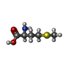
#35: Chemical ChemComp-MET / METHIONINE


Type: L-peptide linking / Mass: 149.211 Da / Num. of mol.: 1 / Source method: obtained synthetically / Formula: C5H11NO2S
#36: Water ChemComp-HOH / water


Mass: 18.015 Da / Num. of mol.: 379 / Source method: isolated from a natural source / Formula: H2O

-
Details

Has ligand of interestN
Has protein modificationN

-
Experimental details

-
Experiment

ExperimentMethod: ELECTRON MICROSCOPY
EM experimentAggregation state: PARTICLE / 3D reconstruction method: single particle reconstruction

-
Sample preparation

ComponentName: 30S ribosomal subunit with initiation factor 1A, mRNA and initiator tRNA
Type: COMPLEX / Entity ID: #1-#32 / Source: NATURAL
Source (natural)Organism: Pyrococcus abyssi GE5 (archaea)
Buffer solutionpH: 6.7
SpecimenEmbedding applied: NO / Shadowing applied: NO / Staining applied: NO / Vitrification applied: YES
Specimen supportGrid material: COPPER / Grid type: Quantifoil R2/1
VitrificationInstrument: LEICA EM GP / Cryogen name: ETHANE

-
Electron microscopy imaging

Experimental equipment
Model: Titan Krios / Image courtesy: FEI Company
MicroscopyModel: TFS KRIOS
Electron gunElectron source: FIELD EMISSION GUN / Accelerating voltage: 300 kV / Illumination mode: FLOOD BEAM
Electron lensMode: BRIGHT FIELD / Nominal defocus max: 2500 nm / Nominal defocus min: 800 nm
Specimen holderSpecimen holder model: FEI TITAN KRIOS AUTOGRID HOLDER
Image recordingElectron dose: 39 e/Å2 / Film or detector model: GATAN K3 BIOQUANTUM (6k x 4k)

-
Processing

SoftwareName: PHENIX / Version: 1.19_4092: / Classification: refinement
EM softwareName: PHENIX / Category: model refinement
CTF correctionType: PHASE FLIPPING AND AMPLITUDE CORRECTION
Particle selectionNum. of particles selected: 2000000
3D reconstructionResolution: 2.6 Å / Resolution method: FSC 0.143 CUT-OFF / Num. of particles: 382130 / Symmetry type: POINT
Atomic model buildingPDB-ID: 7ZHG
Accession code: 7ZHG / Source name: PDB / Type: experimental model
Refine LS restraints
Refine-IDTypeDev idealNumber
ELECTRON MICROSCOPYf_bond_d0.00368832
ELECTRON MICROSCOPYf_angle_d0.534100783
ELECTRON MICROSCOPYf_dihedral_angle_d12.91331108
ELECTRON MICROSCOPYf_chiral_restr0.03712329
ELECTRON MICROSCOPYf_plane_restr0.0046818

+
About Yorodumi

-
News

-
Feb 9, 2022. New format data for meta-information of EMDB entries

New format data for meta-information of EMDB entries

  • Version 3 of the EMDB header file is now the official format.
  • The previous official version 1.9 will be removed from the archive.

Related info.:EMDB header

External links:wwPDB to switch to version 3 of the EMDB data model

-
Aug 12, 2020. Covid-19 info

Covid-19 info

URL: https://pdbj.org/emnavi/covid19.php

New page: Covid-19 featured information page in EM Navigator.

Related info.:Covid-19 info / Mar 5, 2020. Novel coronavirus structure data

+
Mar 5, 2020. Novel coronavirus structure data

Novel coronavirus structure data

Related info.:Yorodumi Speices / Aug 12, 2020. Covid-19 info

External links:COVID-19 featured content - PDBj / Molecule of the Month (242):Coronavirus Proteases

+
Jan 31, 2019. EMDB accession codes are about to change! (news from PDBe EMDB page)

EMDB accession codes are about to change! (news from PDBe EMDB page)

  • The allocation of 4 digits for EMDB accession codes will soon come to an end. Whilst these codes will remain in use, new EMDB accession codes will include an additional digit and will expand incrementally as the available range of codes is exhausted. The current 4-digit format prefixed with “EMD-” (i.e. EMD-XXXX) will advance to a 5-digit format (i.e. EMD-XXXXX), and so on. It is currently estimated that the 4-digit codes will be depleted around Spring 2019, at which point the 5-digit format will come into force.
  • The EM Navigator/Yorodumi systems omit the EMD- prefix.

Related info.:Q: What is EMD? / ID/Accession-code notation in Yorodumi/EM Navigator

External links:EMDB Accession Codes are Changing Soon! / Contact to PDBj

+
Jul 12, 2017. Major update of PDB

Major update of PDB

  • wwPDB released updated PDB data conforming to the new PDBx/mmCIF dictionary.
  • This is a major update changing the version number from 4 to 5, and with Remediation, in which all the entries are updated.
  • In this update, many items about electron microscopy experimental information are reorganized (e.g. em_software).
  • Now, EM Navigator and Yorodumi are based on the updated data.

External links:wwPDB Remediation / Enriched Model Files Conforming to OneDep Data Standards Now Available in the PDB FTP Archive

-
Yorodumi

Thousand views of thousand structures

  • Yorodumi is a browser for structure data from EMDB, PDB, SASBDB, etc.
  • This page is also the successor to EM Navigator detail page, and also detail information page/front-end page for Omokage search.
  • The word "yorodu" (or yorozu) is an old Japanese word meaning "ten thousand". "mi" (miru) is to see.

Related info.:EMDB / PDB / SASBDB / Comparison of 3 databanks / Yorodumi Search / Aug 31, 2016. New EM Navigator & Yorodumi / Yorodumi Papers / Jmol/JSmol / Function and homology information / Changes in new EM Navigator and Yorodumi

Read more